Explore
 Validate
 Learn
Learn Western blot
 ELISA
ELISAAntibody data
- Antibody Data
- Antigen structure
- References [1]
- Comments [0]
- Validations
- Western blot [1]
- Immunocytochemistry [1]
- Immunohistochemistry [1]
- Flow cytometry [1]
Submit
Validation data
Reference
Comment
Report error
- Product number
- NBP1-06014 - Provider product page

- Provider
- Novus Biologicals
- Proper citation
- Novus Cat#NBP1-06014, RRID:AB_1556159
- Product name
- Goat Polyclonal AIF-1/Iba1 Antibody
- Antibody type
- Polyclonal
- Description
- Immunogen affinity purified. This AIF-1/Iba1 Antibody is expected to recognise isoform 1 (NP_116573.1) and isoform 3 (NP_001614.3).
- Reactivity
- Human
- Host
- Goat
- Antigen sequence
C-YEEKAREKEKP- Isotype
- IgG
- Vial size
- 0.1 mg
- Concentration
- 0.5 mg/ml
- Storage
- Store at -20C. Avoid freeze-thaw cycles.
Submitted references Expression of allograft inflammatory factor-1 in T lymphocytes: a role in T-lymphocyte activation and proliferative arteriopathies.
Kelemen SE, Autieri MV
The American journal of pathology 2005 Aug;167(2):619-26
The American journal of pathology 2005 Aug;167(2):619-26
No comments: Submit comment
Supportive validation
- Submitted by
- Novus Biologicals (provider)
- Main image
- Experimental details
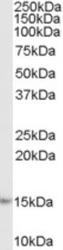
- Western Blot: AIF-1/Iba1 Antibody [NBP1-06014] - (1ug/ml) staining of Human Lymph Node lysate (35ug protein in RIPA buffer). Primary incubation was 1 hour. Detected by chemiluminescence.
Supportive validation
- Submitted by
- Novus Biologicals (provider)
- Main image

- Experimental details
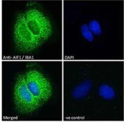
- Immunocytochemistry/Immunofluorescence
Supportive validation
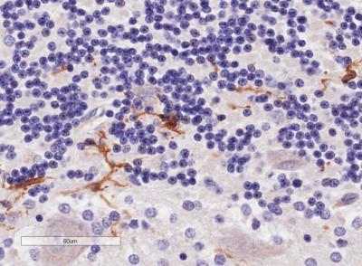
- Submitted by
- Novus Biologicals (provider)
- Main image
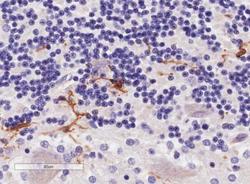
- Experimental details
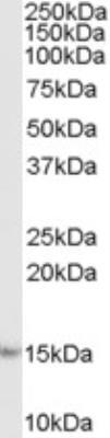
- Immunohistochemistry-Paraffin: AIF-1/Iba1 Antibody [NBP1-06014] - Staining of paraffin embedded Human Cerebellum AIF-1/Iba1 Antibody at 4 ug/mL. Microwaved antigen retrieval with Tris/EDTA buffer pH9, HRP-staining.
Supportive validation
- Submitted by
- Novus Biologicals (provider)
- Main image
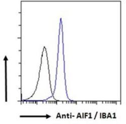
- Experimental details
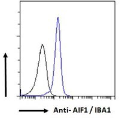
- Flow Cytometry: AIF-1/Iba1 Antibody [NBP1-06014] - Flow cytometric analysis of paraformaldehyde fixed K562 cells (blue line), permeabilized with 0.5% Triton. Primary incubation 1hr (10 ug/mL) with AIF-1:Iba1 Antibody, followed by Alexa Fluor 488 secondary antibody (1 ug/mL). IgG control: Unimmunized goat IgG (black line) followed by Alexa Fluor 488 secondary antibody.