Explore
 Validate
 Learn
Learn Western blot
 Immunohistochemistry
Antibody data
- Antibody Data
- Antigen structure
- References [6]
- Comments [0]
- Validations
- Immunohistochemistry [3]
- Chromatin Immunoprecipitation [2]
- Other assay [8]
Submit
Validation data
Reference
Comment
Report error
- Product number
- PA5-40591 - Provider product page

- Provider
- Invitrogen Antibodies
- Product name
- KLF2 Polyclonal Antibody
- Antibody type
- Polyclonal
- Antigen
- Synthetic peptide
- Description
- Peptide sequence: PPAFGLFDDA AAAAAALGLA PPAARGLLTP PASPLELLEA KPKRGRRSWP Sequence homology: Guinea Pig: 93%; Human: 100%; Mouse: 93%; Pig: 100%; Rat: 93%; Zebrafish: 79%
- Reactivity
- Human
- Host
- Rabbit
- Isotype
- IgG
- Vial size
- 100 μL
- Concentration
- 0.5 mg/mL
- Storage
- -20°C, Avoid Freeze/Thaw Cycles
Submitted references Intermittent High Glucose Elevates Nuclear Localization of EZH2 to Cause H3K27me3-Dependent Repression of KLF2 Leading to Endothelial Inflammation.
IRF2BP2 prevents ox-LDL-induced inflammation and EMT in endothelial cells via regulation of KLF2.
MiR-144-induced KLF2 inhibition and NF-kappaB/CXCR1 activation promote neutrophil extracellular trap-induced transfusion-related acute lung injury.
Retinoblastoma cell-derived exosomes promote angiogenesis of human vesicle endothelial cells through microRNA-92a-3p.
Diagnostic Utility and Pathogenic Role of Circulating MicroRNAs in Vasospastic Angina.
Sex differences in the proliferation of pulmonary artery endothelial cells: implications for plexiform arteriopathy.
Thakar S, Katakia YT, Ramakrishnan SK, Pandya Thakkar N, Majumder S
Cells 2021 Sep 26;10(10)
Cells 2021 Sep 26;10(10)
IRF2BP2 prevents ox-LDL-induced inflammation and EMT in endothelial cells via regulation of KLF2.
Jiang Y, Shen Q
Experimental and therapeutic medicine 2021 May;21(5):481
Experimental and therapeutic medicine 2021 May;21(5):481
MiR-144-induced KLF2 inhibition and NF-kappaB/CXCR1 activation promote neutrophil extracellular trap-induced transfusion-related acute lung injury.
Le A, Wu Y, Liu W, Wu C, Hu P, Zou J, Kuang L
Journal of cellular and molecular medicine 2021 Jul;25(14):6511-6523
Journal of cellular and molecular medicine 2021 Jul;25(14):6511-6523
Retinoblastoma cell-derived exosomes promote angiogenesis of human vesicle endothelial cells through microRNA-92a-3p.
Chen S, Chen X, Luo Q, Liu X, Wang X, Cui Z, He A, He S, Jiang Z, Wu N, Chen P, Yu K, Zhuang J
Cell death & disease 2021 Jul 13;12(7):695
Cell death & disease 2021 Jul 13;12(7):695
Diagnostic Utility and Pathogenic Role of Circulating MicroRNAs in Vasospastic Angina.
Park CS, Kim I, Oh GC, Han JK, Yang HM, Park KW, Cho HJ, Kang HJ, Koo BK, Chung WY, Oh S, Lee HY
Journal of clinical medicine 2020 May 2;9(5)
Journal of clinical medicine 2020 May 2;9(5)
Sex differences in the proliferation of pulmonary artery endothelial cells: implications for plexiform arteriopathy.
Qin S, Predescu DN, Patel M, Drazkowski P, Ganesh B, Predescu SA
Journal of cell science 2020 May 14;133(9)
Journal of cell science 2020 May 14;133(9)
No comments: Submit comment
Supportive validation
- Submitted by
- Invitrogen Antibodies (provider)
- Main image
- Experimental details
- Immunohistochemistry analysis of human tissue using an anti-KLF2 polyclonal antibody (Product # PA5-40591).
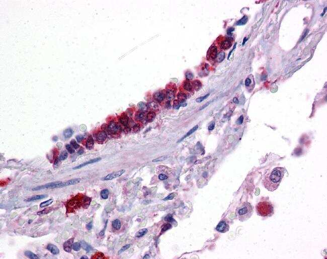
- Submitted by
- Invitrogen Antibodies (provider)
- Main image

- Experimental details
- Immunohistochemistry analysis of human lung tissue using an anti-KLF2 polyclonal antibody (Product # PA5-40591).
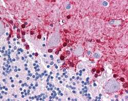
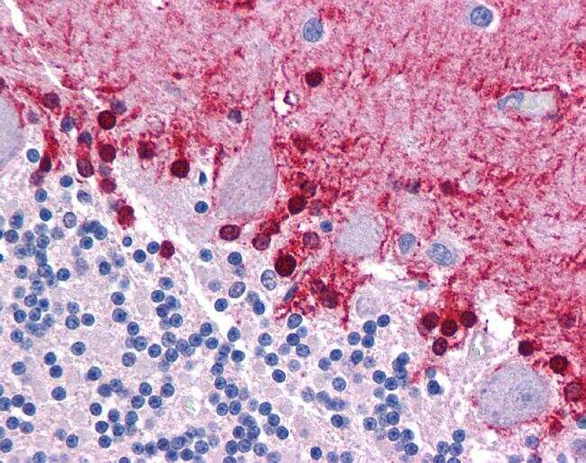
- Submitted by
- Invitrogen Antibodies (provider)
- Main image
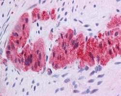
- Experimental details
- Immunohistochemistry analysis of human brain tissue using an anti-KLF2 polyclonal antibody (Product # PA5-40591).

Supportive validation
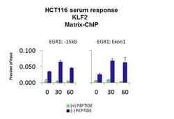
- Submitted by
- Invitrogen Antibodies (provider)
- Main image

- Experimental details
- ChIP assay analysis of quiescent human colon carcinoma HCT116 cells using an anti-KLF2 polyclonal antibody (Product # PA5-40591).

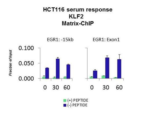
- Submitted by
- Invitrogen Antibodies (provider)
- Main image
- Experimental details
- ChIP assay analysis of quiescent human colon carcinoma HCT116 cells using an anti-KLF2 polyclonal antibody (Product # PA5-40591).
Supportive validation
- Submitted by
- Invitrogen Antibodies (provider)
- Main image
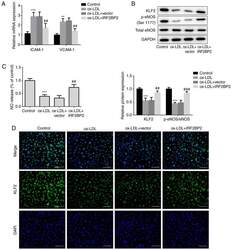
- Experimental details
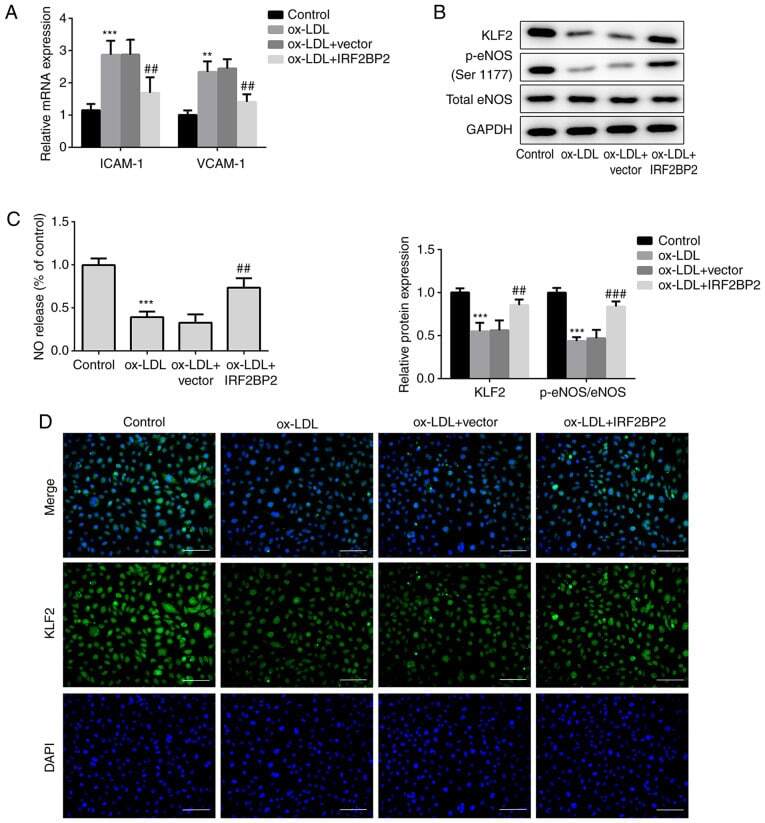
- Figure 2 IRF2BP2 increases the expression of KLF2 and improves endothelial function. (A) The expression levels of ICAM-1 and VCAM-1 were determined using reverse transcription-quantitative PCR analysis. (B) The protein expression levels of KLF2, p-eNOS, total eNOS and GAPDH were measured using western blot analysis. (C) A Nitrate/Nitrite assay kit was utilized to estimate the release of NO. (D) The expression of KLF2 was evaluated by immunofluorescence analysis (magnification, x200). Scale bar, 50 um. ** P
- Submitted by
- Invitrogen Antibodies (provider)
- Main image
- Experimental details
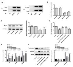
- Figure 3 IRF2BP2 attenuates inflammation and EMT via KLF2. (A) The interaction between IRF2BP2 and KLF2 was confirmed using a Co-IP assay. (B) HUVECs were transfected with scrambled siRNA, siKLF2#1 or siKLF2#2. The mRNA expression levels of KLF2 were measured using reverse transcription-quantitative PCR analysis. *** P
- Submitted by
- Invitrogen Antibodies (provider)
- Main image

- Experimental details
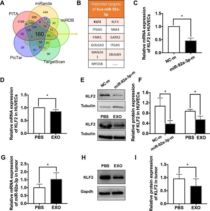
- Fig. 7 Exosomal delivered miR-92a-3p modulates angiogenesis by targeting KLF2. A Venn diagram showing overlap of the data from miRanda, miRDB, TargetScan, PicTar, and PITA. B Potential targets of hsa-miR-92a-3p. C The mRNA level of KLF2 in the HUVECs transfected with synthetic mimic control (NC-m) or miR-92a-3p mimic (miR-92a-3p-m) ( n = 4, * P < 0.05). D The mRNA level of KLF2 in the HUVECs treated with PBS or EXOs ( n = 5, * P < 0.05). E Western blot analysis and relative quantification of protein expression indicating that exosome treatment or overexpression of miR-92a-3p could downregulate KLF2 expression in HUVECs ( n >= 6, * P < 0.05). G The mRNA level of miR-92a-3p in the tumor tissues treated with PBS or exosomes ( n = 6, * P < 0.05). H Representative blots of KLF2 in the tumor tissues treated with PBS or exosomes. I Quantitative evaluation of KLF2 protein expression in the tumor tissues treated with PBS or exosomes ( n = 6, * P < 0.05).
- Submitted by
- Invitrogen Antibodies (provider)
- Main image
- Experimental details
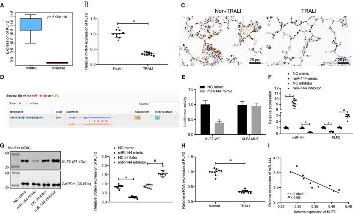
- FIGURE 3 miR-144 directly targets and negatively regulates KLF2. A, Box plots displaying the expression of KLF2 in normal samples (blue box on the left) and TRALI samples (red box on the right) from microarray data set GSE11434 . B, The mRNA level of KLF2 in blood samples from healthy people and patients with TRALI determined by RT-qPCR. C, Immunohistochemistry to detect the level of KLF2 in lung tissues of patients with TRALI (x400). D, The putative binding sites between miR-144 and KLF2 predicted from the Starbase website. E, The binding relationship between miR-144 and KLF2 verified by a dual-luciferase reporter gene assay. F, The mRNA levels of miR-144 and KLF2 in human blood samples after transfection of miR-144 mimic/inhibitor measured by RT-qPCR. G, KLF2 protein levels in human blood samples after transfection of miR-144 mimic/inhibitor normalized to GAPDH evaluated by Western blot analysis. H, KLF2 mRNA levels in normal and TRALI mouse blood samples after transfection examined by RT-qPCR. I, Pearson correlation analysis of the correlation between miR-144 and KLF2 mRNA expression in mice. The measurement data were expressed by the mean +- standard deviation. The experiment was repeated 3 times independently. The comparison between the two groups was analysed by an unpaired t test. Data among multiple groups were compared using a one-way ANOVA, followed by Tukey's post hoc test. Pearson correlation was used for correlation analysis. * P < .05 vs. healthy donors, the NC
- Submitted by
- Invitrogen Antibodies (provider)
- Main image
- Experimental details
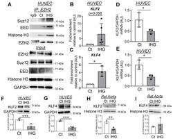
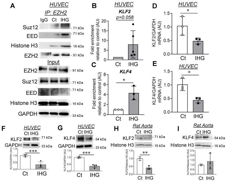
- Figure 3 Intermittent hyperglycemia causes EZH2 coupling with PRC2 proteins, leading to H3K27 trimethylation at promoter regions of the KLF2 and KLF4 genes. ( A ) Co-immunoprecipitation with EZH2 antibody, followed by immunoblotting for Suz12, EED, and histone H3 in both immuno-precipitated and total cell lysate (input) sample ( n = 3). ( B , C ) ChIP assay using H3K27me3 antibody, followed by qPCR using primers specific to amplify promoter regions of KLF2 gene (( B ), n = 5) and KLF4 gene (( C ), n = 3) in HUVEC cells treated with intermittent high glucose. ( D , E ) Transcriptomic quantitation of KLF2 (( D ), n = 3) and KLF4 (( E ), n = 3) in HUVEC challenged with intermittent high glucose. ( F , G ) Immunoblotting and quantitation for KLF2 (( F ), n = 4) and KLF4 (( G ), n = 4) in intermittent high glucose-challenged HUVEC. ( H , I ). Tissue lysates of intermittent high glucose-treated rat aortic rings were immunoblotted for KLF2 (( H ), n = 3) and KLF4 (( I ), n = 3). Control treatment condition (cells constantly exposed to normal glucose (5.5 mM)) represented as ""Ct"" in the figure. All analyzed data were normalized to the control treatment condition. Values represent the mean +- SD. * p < 0.05, ** p < 0.01, and *** p < 0.001 by unpaired t test.
- Submitted by
- Invitrogen Antibodies (provider)
- Main image
- Experimental details
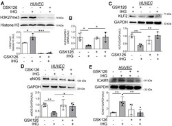
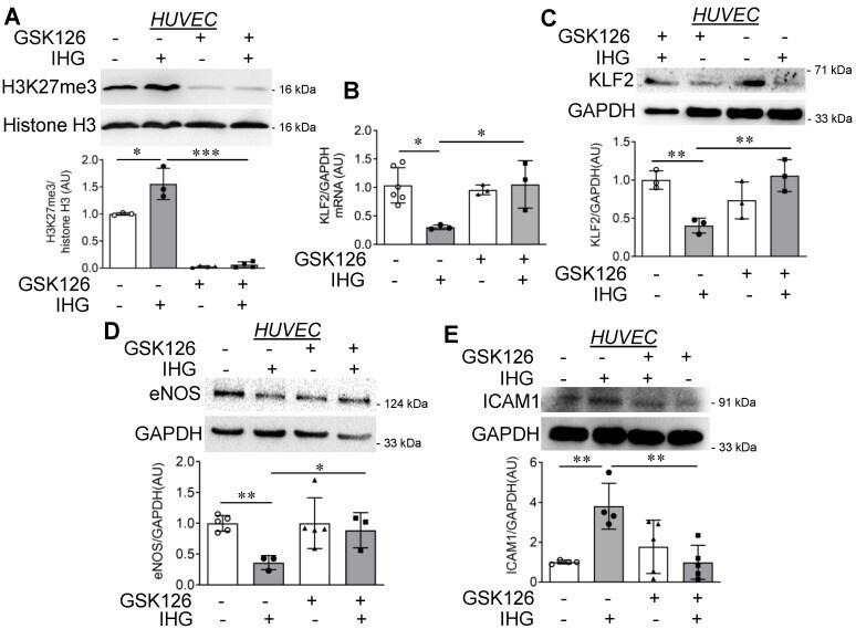
- Figure 4 Small molecule-mediated inhibition of EZH2 in intermittent high glucose-exposed HUVEC decreases H3K27me3 level and blocks the inflammatory switch of EC. ( A ) Immunoblotting for H3K27me3 ( n = 3) in GSK126-treated and intermittent high glucose-exposed HUVEC. ( B ) KLF2 mRNA ( n = 3) expression in HUVEC challenged with intermittent high glucose (alternating 5.5 mM (normal glucose) and 25 mM (high glucose) cycle of 12 h for 3 cycles) in combination with GSK126 (10 muM for 72 h). ( C - E ) KLF2 ( C ), eNOS ( D ), and ICAM1 ( E ) in cultured HUVEC treated with the EZH2 inhibitor GSK126 (10 muM for 72 h) in combination with intermittent high glucose ( n = 3). Cells continuously exposed to normal glucose (5.5 mM) were considered as the non-intermittent high glucose treatment condition. All analyzed data were normalized to the control treatment condition. Values represent the mean +- SD. * p < 0.05, ** p < 0.01, and *** p < 0.001 by unpaired t test.
- Submitted by
- Invitrogen Antibodies (provider)
- Main image
- Experimental details
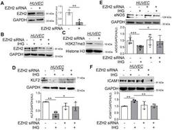
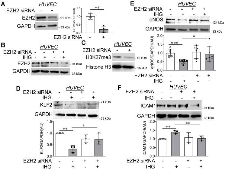
- Figure 5 EZH2 knockdown in HUVEC reduces H3K27me3 and reverses intermittent high glucose-driven endothelial inflammation. ( A - E ) Immunoblotting of EZH2 in normal glucose (5.5 mM)-treated HUVEC (( A ), n = 4), EZH2 in both normal glucose (5.5 mM) and intermittent high glucose-treated HUVEC (( B ), n = 3), H3K27me3 (( C ), n = 3) and quantification of KLF2 (( D ), n = 3), eNOS (( E ), n = 6), and ICAM1 (( F ), n = 3) in HUVEC cells transiently transfected with scrambled or EZH2 siRNA, followed by challenging with intermittent high glucose. Cells continuously exposed to normal glucose (5.5 mM) were considered as the non-intermittent high glucose treatment condition. All analyzed data were normalized to the control treatment condition. Values represent the mean +- SD. * p < 0.05, ** p < 0.01, and *** p < 0.001 by unpaired t test.
- Submitted by
- Invitrogen Antibodies (provider)
- Main image
- Experimental details
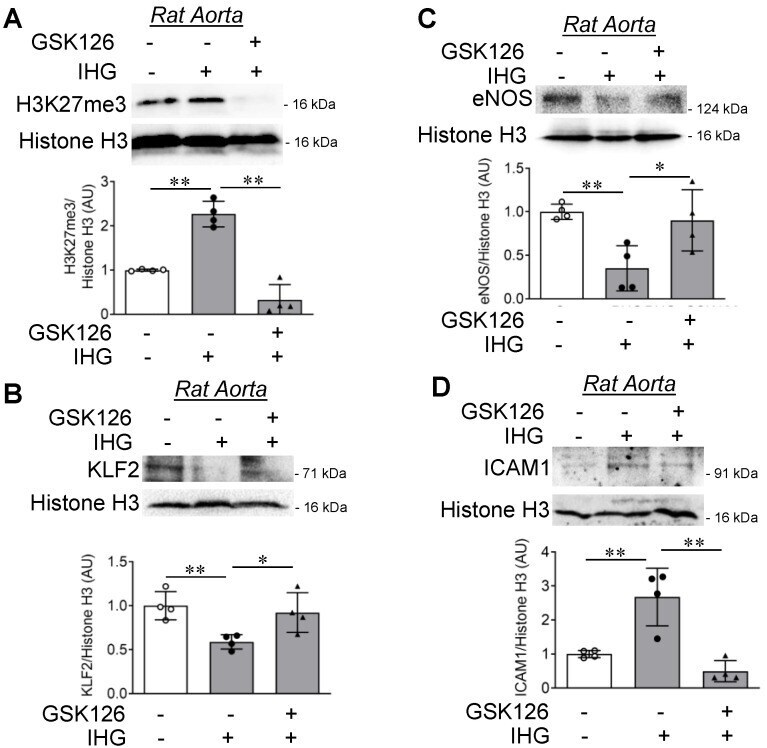
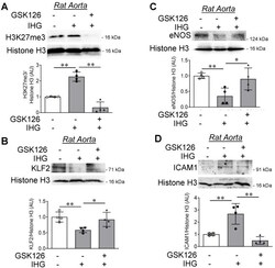
- Figure 6 Inhibition of EZH2 in rat aortic rings exposed to intermittent hyperglycemia reduces H3K27me3 levels and blocks inflammatory signaling ex vivo. ( A - D ) Immunoblotting for H3K27me3 (( A ), n = 4), KLF2 (( B ), n = 4), eNOS (( C ), n = 4), and ICAM1 (( D ), n = 4) in rat aortic rings challenged with the EZH2 inhibitor GSK126 (10 muM for 72 h) in combination with intermittent high glucose. Tissues continuously exposed to normal glucose (5.5 mM) were considered as the non-intermittent high glucose treatment condition. All analyzed data were normalized to the control treatment condition. Values represent the mean +- SD. * p < 0.05 and ** p < 0.01 by unpaired t test.