Explore
 Validate
 Learn
Learn Western blot
 Flow cytometry
Antibody data
- Antibody Data
- Antigen structure
- References [0]
- Comments [0]
- Validations
- Flow cytometry [1]
Submit
Validation data
Reference
Comment
Report error
- Product number
- MAB1022 - Provider product page

- Provider
- R&D Systems
- Product name
- Mouse PD-L2/B7-DC Antibody
- Antibody type
- Monoclonal
- Description
- Protein A or G purified from hybridoma culture supernatant. Detects mouse PD-L2/B7-DC in direct ELISAs and Western blots. In direct ELISAs and Western blots, 100% cross-reactivity with recombinant human (rh) PD-L2 is observed and no cross-reactivity with rhB7-1, recombinant mouse (rm) B7-1, rmB7-2, rmB7-H1, rmB7-H2, rmB7-H3, or rmB7-H4 is observed.
- Reactivity
- Mouse
- Host
- Rat
- Conjugate
- Unconjugated
- Antigen sequence
Q9WUL5- Isotype
- IgG
- Antibody clone number
- 168633
- Vial size
- 100 ug
- Concentration
- LYOPH
- Storage
- Use a manual defrost freezer and avoid repeated freeze-thaw cycles. 12 months from date of receipt, -20 to -70 °C as supplied. 1 month, 2 to 8 °C under sterile conditions after reconstitution. 6 months, -20 to -70 °C under sterile conditions after reconstitution.
No comments: Submit comment
Supportive validation
- Submitted by
- R&D Systems (provider)
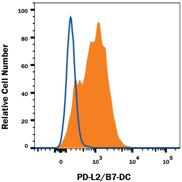
- Main image

- Experimental details
- Detection of PD-L2/B7-DC in HEK293 Human Cell Line Transfected with Mouse PD-L2/B7-DC by Flow Cytometry. HEK293 human embryonic kidney cell line transfected with either mouse PD-L2/B7-DC (filled histogram) or irrelevant transfectants (open histogram) was stained with Rat Anti-Mouse PD-L2/B7-DC Monoclonal Antibody (Catalog # MAB1022), followed by Allophycocyanin-conjugated Anti-Rat IgG Secondary Antibody (Catalog # F0113). View our protocol for Staining Membrane-associated Proteins.