Explore
 Validate
 Learn
LearnNBP1-76770
antibody from Novus Biologicals
Targeting: PDCD1LG2
B7-DC, bA574F11.2, Btdc, CD273, PD-L2, PDL2
 Western blot
 ELISA
ELISAAntibody data
- Antibody Data
- Antigen structure
- References [0]
- Comments [0]
- Validations
- Western blot [5]
- Immunohistochemistry [3]
Submit
Validation data
Reference
Comment
Report error
- Product number
- NBP1-76770 - Provider product page

- Provider
- Novus Biologicals
- Proper citation
- Novus Cat#NBP1-76770, RRID:AB_11009464
- Product name
- Rabbit Polyclonal PD-L2/B7-DC/PDCD1LG2 Antibody
- Antibody type
- Polyclonal
- Description
- Peptide affinity purified.
- Reactivity
- Human, Mouse, Rat
- Host
- Rabbit
- Isotype
- IgG
- Vial size
- 0.1 mg
- Concentration
- 1 mg/ml
- Storage
- Store at 4C short term. Aliquot and store at -20C long term. Avoid freeze-thaw cycles.
No comments: Submit comment
Supportive validation
- Submitted by
- Novus Biologicals (provider)
- Main image

- Experimental details
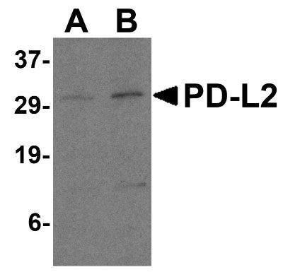
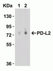
- Western Blot: PD-L2/B7-DC/PDCD1LG2 Antibody [NBP1-76770] - Analysis of PD-L2 in Raji cell lysate with PD-L2 antibody at 0.5 and 1 ug/mL.
- Submitted by
- Novus Biologicals (provider)
- Main image

- Experimental details
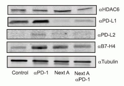
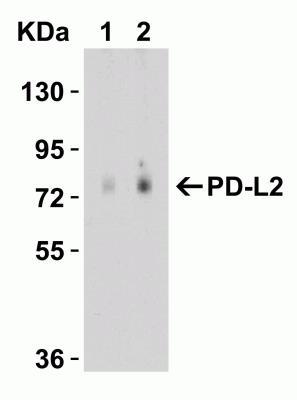
- Western Blot: PD-L2/B7-DC/PDCD1LG2 Antibody [NBP1-76770] - PD-L2 expression was up-regulated by anti-PD1 antibody treatment whereas it was reduced by Next A alone or combination treatment (anti-PD1 antibody+NextA).
- Submitted by
- Novus Biologicals (provider)
- Main image

- Experimental details
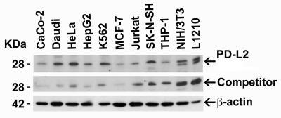
- Western Blot: PD-L2/B7-DC/PDCD1LG2 Antibody [NBP1-76770] - Loading: 15 ug of lysates per lane. Antibodies: PD-L2 (4 ug/mL), competitor antibody (4 ug/mL), and beta-actin (1 ug/mL), 1h incubation at RT in 5% NFDM/TBST. Secondary: Goat anti-rabbit IgG HRP conjugate at 1:10000 dilution.
- Submitted by
- Novus Biologicals (provider)
- Main image

- Experimental details
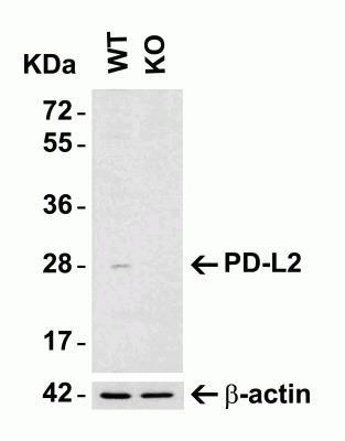
- Western Blot: PD-L2/B7-DC/PDCD1LG2 Antibody [NBP1-76770] - Loading: 15 ug of HeLa WT cell lysates or PD-L2 KO cell lysates. Antibodies: PD-L2, 4063 (4 ug/mL) and beta-actin (1 ug/mL), 1 h incubation at RT in 5% NFDM/TBST. Secondary: Goat Anti-Rabbit IgG HRP conjugate at 1:10000 dilution
- Submitted by
- Novus Biologicals (provider)
- Main image
- Experimental details
- Western Blot: PD-L2/B7-DC/PDCD1LG2 Antibody [NBP1-76770] - Loading: 15 ug of HeLa WT cell lysates or PD-L2 KO cell lysates. Antibodies: PD-L2, 4063 (4 ug/mL) and beta-actin (1 ug/mL), 1 h incubation at RT in 5% NFDM/TBST. Secondary: Goat Anti-Rabbit IgG HRP conjugate at 1:10000 dilution.
Supportive validation
- Submitted by
- Novus Biologicals (provider)
- Main image

- Experimental details
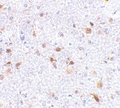
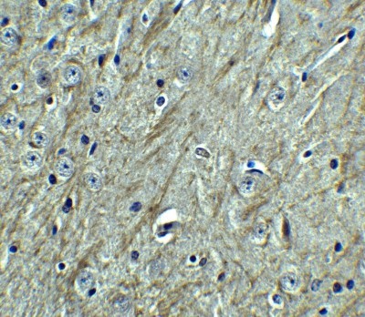
- Immunohistochemistry-Paraffin: PD-L2/B7-DC/PDCD1LG2 Antibody [NBP1-76770] - Mouse brain tissue.
- Submitted by
- Novus Biologicals (provider)
- Main image

- Experimental details
- Immunohistochemistry: PD-L2/B7-DC/PDCD1LG2 Antibody [NBP1-76770] - Staining of mouse brain tissue with antibody at 2.5 ug/ml.
- Submitted by
- Novus Biologicals (provider)
- Main image

- Experimental details
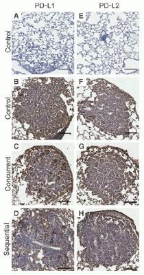
- Immunohistochemistry-Paraffin: PD-L2/B7-DC/PDCD1LG2 Antibody [NBP1-76770] - Protein analysis for PD-L2 (E-H) by immunohistochemistry with anti-PD-L2 antibodies in mice lung tumors. hMUC1.Tg mice were induced with lung adenoma and then treated with concurrent or sequential cisplatin/radiotherapy. PDL2 expression level at week 41 after treatment was similar in control and treatment groups.