Explore
 Validate
 Learn
Learn Western blot
 ELISA
ELISAAntibody data
- Antibody Data
- Antigen structure
- References [9]
- Comments [0]
- Validations
- Western blot [1]
- Immunohistochemistry [1]
Submit
Validation data
Reference
Comment
Report error
- Product number
- 11100-2-AP - Provider product page

- Provider
- Proteintech Group
- Proper citation
- Proteintech Cat#11100-2-AP, RRID:AB_2289959
- Product name
- Arrestin C antibody
- Antibody type
- Polyclonal
- Description
- Arrestin C antibody (Cat. #11100-2-AP) is a rabbit polyclonal antibody that shows reactivity with human, mouse and has been validated for the following applications: IHC, WB, ELISA.
- Reactivity
- Human, Mouse
- Host
- Rabbit
- Conjugate
- Unconjugated
- Isotype
- IgG
- Vial size
- 20ul, 150ul
Submitted references Spatial transcriptomics of retinoblastoma: a visual window on intra-patient heterogeneity.
Clinical features and molecular mechanisms of RP1L1 variants causing occult macular dystrophy.
Targeting ZIP8 mediated ferroptosis as a novel strategy to protect against the retinal pigment epithelial degeneration.
Immunohistochemical expression of TFF1 is a marker of poor prognosis in retinoblastoma.
Establishment and Comprehensive Characterization of a Novel Preclinical Platform of Metastatic Retinoblastoma for Therapeutic Developments.
A mouse model of cone photoreceptor function loss (cpfl9) with degeneration due to a mutation in Gucy2e.
A high-risk retinoblastoma subtype with stemness features, dedifferentiated cone states and neuronal/ganglion cell gene expression.
Clinical, Genomic, and Pharmacological Study of MYCN-Amplified RB1 Wild-Type Metastatic Retinoblastoma.
Tridimensional Retinoblastoma Cultures as Vitreous Seeds Models for Live-Cell Imaging of Chemotherapy Penetration.
Moulin AP, Thevenet J, Mazzeo L, Tissot S, Stathopoulos C, Munier FL, Berger A
BMC cancer 2025 Sep 2;25(1):1410
BMC cancer 2025 Sep 2;25(1):1410
Clinical features and molecular mechanisms of RP1L1 variants causing occult macular dystrophy.
Pan Y, Iejima D, Yoshitake K, Tsunoda K, Iwata T, Japan Eye Genetics Consortium
HGG advances 2025 Jul 10;6(3):100461
HGG advances 2025 Jul 10;6(3):100461
Targeting ZIP8 mediated ferroptosis as a novel strategy to protect against the retinal pigment epithelial degeneration.
Liu Z, Huang J, Li D, Zhang C, Wan H, Zeng B, Tan Y, Zhong F, Liao H, Liu M, Chen ZS, Zou C, Liu D, Qin B
Free radical biology & medicine 2024 Mar;214:42-53
Free radical biology & medicine 2024 Mar;214:42-53
Immunohistochemical expression of TFF1 is a marker of poor prognosis in retinoblastoma.
Aschero R, Ganiewich D, Lamas G, Restrepo-Perdomo CA, Ottaviani D, Zugbi S, Camarero S, Néspoli E, Vilanova MC, Perez-Jaume S, Pascual-Pasto G, Sampor C, Grigorovski N, Salas B, Suñol M, Carcaboso AM, Mora J, de Dávila MTG, Doz F, Radvanyi F, Abramson DH, Llera AS, Schaiquevich PS, Lubieniecki F, Chantada GL
Pediatric blood & cancer 2024 Jan;71(1):e30717
Pediatric blood & cancer 2024 Jan;71(1):e30717
Establishment and Comprehensive Characterization of a Novel Preclinical Platform of Metastatic Retinoblastoma for Therapeutic Developments.
Zugbi S, Aschero R, Ganiewich D, Cancela MB, Winter U, Ottaviani D, Sampor C, Dinardi M, Torbidoni AV, Mena M, Balaguer-Lluna L, Lamas G, Sgroi M, Lagomarsino E, Lubieniecki F, Fandiño A, Radvanyi F, Abramson DH, Podhajcer O, Llera AS, Cafferata EG, Chantada G, Carcaboso AM, Schaiquevich P
Investigative ophthalmology & visual science 2023 Dec 1;64(15):27
Investigative ophthalmology & visual science 2023 Dec 1;64(15):27
A mouse model of cone photoreceptor function loss (cpfl9) with degeneration due to a mutation in Gucy2e.
Naggert ASEN, Collin GB, Wang J, Krebs MP, Chang B
Frontiers in molecular neuroscience 2022;15:1080136
Frontiers in molecular neuroscience 2022;15:1080136
A high-risk retinoblastoma subtype with stemness features, dedifferentiated cone states and neuronal/ganglion cell gene expression.
Liu J, Ottaviani D, Sefta M, Desbrousses C, Chapeaublanc E, Aschero R, Sirab N, Lubieniecki F, Lamas G, Tonon L, Dehainault C, Hua C, Fréneaux P, Reichman S, Karboul N, Biton A, Mirabal-Ortega L, Larcher M, Brulard C, Arrufat S, Nicolas A, Elarouci N, Popova T, Némati F, Decaudin D, Gentien D, Baulande S, Mariani O, Dufour F, Guibert S, Vallot C, Rouic LL, Matet A, Desjardins L, Pascual-Pasto G, Suñol M, Catala-Mora J, Llano GC, Couturier J, Barillot E, Schaiquevich P, Gauthier-Villars M, Stoppa-Lyonnet D, Golmard L, Houdayer C, Brisse H, Bernard-Pierrot I, Letouzé E, Viari A, Saule S, Sastre-Garau X, Doz F, Carcaboso AM, Cassoux N, Pouponnot C, Goureau O, Chantada G, de Reyniès A, Aerts I, Radvanyi F
Nature communications 2021 Sep 22;12(1):5578
Nature communications 2021 Sep 22;12(1):5578
Clinical, Genomic, and Pharmacological Study of MYCN-Amplified RB1 Wild-Type Metastatic Retinoblastoma.
Zugbi S, Ganiewich D, Bhattacharyya A, Aschero R, Ottaviani D, Sampor C, Cafferata EG, Mena M, Sgroi M, Winter U, Lamas G, Suñol M, Daroqui M, Baialardo E, Salas B, Das A, Fandiño A, Francis JH, Lubieniecki F, Lavarino C, Garippa R, Podhajcer OL, Abramson DH, Radvanyi F, Chantada G, Llera AS, Schaiquevich P
Cancers 2020 Sep 22;12(9)
Cancers 2020 Sep 22;12(9)
Tridimensional Retinoblastoma Cultures as Vitreous Seeds Models for Live-Cell Imaging of Chemotherapy Penetration.
Winter U, Aschero R, Fuentes F, Buontempo F, Zugbi S, Sgroi M, Sampor C, Abramson DH, Carcaboso AM, Schaiquevich P
International journal of molecular sciences 2019 Mar 2;20(5)
International journal of molecular sciences 2019 Mar 2;20(5)
No comments: Submit comment
Supportive validation
- Submitted by
- Proteintech Group (provider)
- Main image
- Experimental details
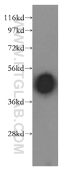
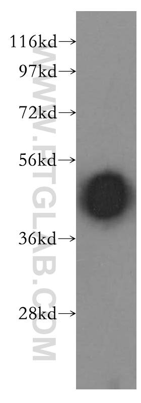
- human brain tissue were subjected to SDS PAGE followed by western blot with 11100-2-AP(ARR3 antibody) at dilution of 1:500
- Sample type
- tissue
Supportive validation
- Submitted by
- Proteintech Group (provider)
- Main image
- Experimental details
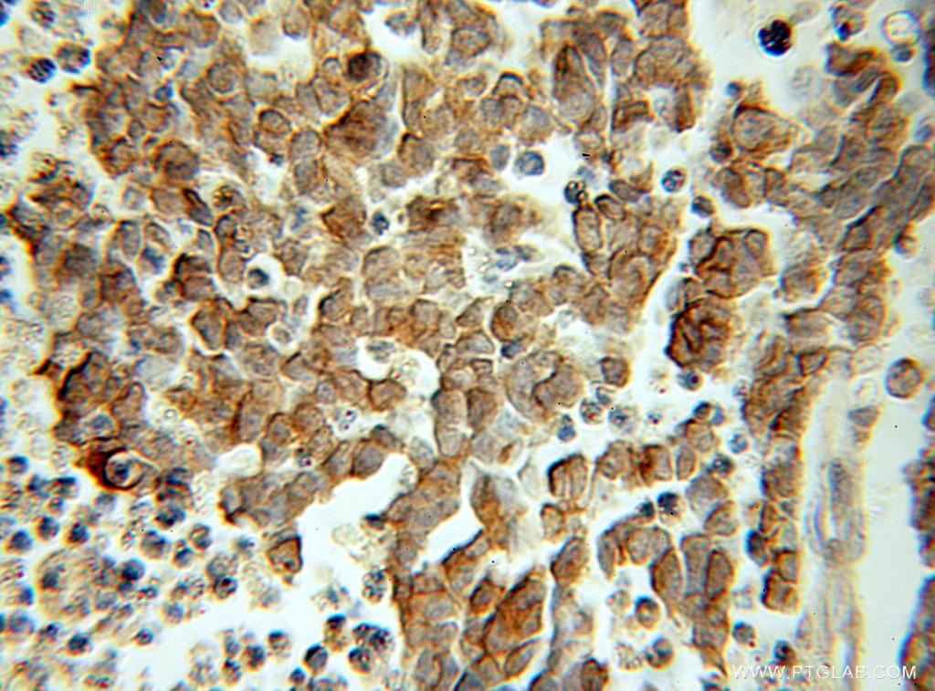
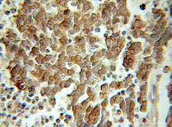
- The Arrestin C antibody from Proteintech is a rabbit polyclonal antibody to a recombinant protein of human Arrestin C. This antibody recognizes human, mouse antigen. The Arrestin C antibody has been validated for the following applications: ELISA, WB, IHC analysis.