Explore
 Validate
 Learn
Learn Western blot
Antibody data
- Antibody Data
- Antigen structure
- References [3]
- Comments [0]
- Validations
- Western blot [1]
- Immunohistochemistry [1]
Submit
Validation data
Reference
Comment
Report error
- Product number
- AF2929 - Provider product page

- Provider
- R&D Systems
- Product name
- Human TIM-4 Antibody
- Antibody type
- Polyclonal
- Description
- Antigen Affinity-purified. Detects human TIM-4 in direct ELISAs and Western blots. In direct ELISAs, less than 1% cross-reactivity with recombinant human (rh) TIM-1 and recombinant mouse (rm) TIM-4 is observed.
- Reactivity
- Human
- Host
- Goat
- Conjugate
- Unconjugated
- Antigen sequence
Q96H15- Isotype
- IgG
- Vial size
- 100 ug
- Concentration
- LYOPH
- Storage
- Use a manual defrost freezer and avoid repeated freeze-thaw cycles. 12 months from date of receipt, -20 to -70 °C as supplied. 1 month, 2 to 8 °C under sterile conditions after reconstitution. 6 months, -20 to -70 °C under sterile conditions after reconstitution.
Submitted references Human Sertoli cells support high levels of Zika virus replication and persistence.
Characterizing functional domains for TIM-mediated enveloped virus entry.
TIM-1 and TIM-4 glycoproteins bind phosphatidylserine and mediate uptake of apoptotic cells.
Kumar A, Jovel J, Lopez-Orozco J, Limonta D, Airo AM, Hou S, Stryapunina I, Fibke C, Moore RB, Hobman TC
Scientific reports 2018 Apr 3;8(1):5477
Scientific reports 2018 Apr 3;8(1):5477
Characterizing functional domains for TIM-mediated enveloped virus entry.
Moller-Tank S, Albritton LM, Rennert PD, Maury W
Journal of virology 2014 Jun;88(12):6702-13
Journal of virology 2014 Jun;88(12):6702-13
TIM-1 and TIM-4 glycoproteins bind phosphatidylserine and mediate uptake of apoptotic cells.
Kobayashi N, Karisola P, Peña-Cruz V, Dorfman DM, Jinushi M, Umetsu SE, Butte MJ, Nagumo H, Chernova I, Zhu B, Sharpe AH, Ito S, Dranoff G, Kaplan GG, Casasnovas JM, Umetsu DT, Dekruyff RH, Freeman GJ
Immunity 2007 Dec;27(6):927-40
Immunity 2007 Dec;27(6):927-40
No comments: Submit comment
Supportive validation
- Submitted by
- R&D Systems (provider)
- Main image

- Experimental details
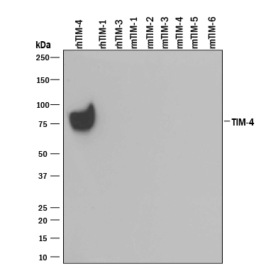
- Detection of Recombinant Human TIM-4 by Western Blot. Western blot shows 25 ng of Recombinant Human TIM-4 His-tag (Catalog # 2929-TM), Recombinant Human TIM-1/KIM-1/HAVCR (Catalog # 1750-TM), Recombinant Human TIM-3 Fc Chimera (Catalog # 2365-TM), Recombinant Mouse TIM-1/KIM-1/HAVCR (Catalog # 1817-TM), Recombinant Mouse TIM-2 (Catalog # 1885-TM), Recombinant Mouse TIM-3 Fc Chimera (Catalog # 1529-TM), Recombinant Mouse TIM-4 (Catalog # 2826-TI), Recombinant Mouse TIM-5, and Recombinant Mouse TIM-6. PVDF Membrane was probed with 0.1 µg/mL of Goat Anti-Human TIM-4 Antigen Affinity-purified Polyclonal Antibody (Catalog # AF2929) followed by HRP-conjugated Anti-Goat IgG Secondary Antibody (Catalog # HAF109). A specific band was detected for TIM-4 at approximately 75 kDa (as indicated). This experiment was conducted under reducing conditions and using Immunoblot Buffer Group 3.
Supportive validation
- Submitted by
- R&D Systems (provider)
- Main image

- Experimental details
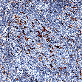
- TIM-4 in Human Tonsil. TIM-4 was detected in immersion fixed paraffin-embedded sections of human tonsil using Goat Anti-Human TIM-4 Antigen Affinity-purified Polyclonal Antibody (Catalog # AF2929) at 1 µg/mL for 1 hour at room temperature followed by incubation with the Anti-Goat IgG VisUCyte™ HRP Polymer Antibody (Catalog # VC004). Before incubation with the primary antibody, tissue was subjected to heat-induced epitope retrieval using Antigen Retrieval Reagent-Basic (Catalog # CTS013). Tissue was stained using DAB (brown) and counterstained with hematoxylin (blue). Specific staining was localized to cell membranes of macrophages. View our protocol for IHC Staining with VisUCyte HRP Polymer Detection Reagents.