Explore
 Validate
 Learn
Learn Western blot
 ELISA
ELISAAntibody data
- Antibody Data
- Antigen structure
- References [12]
- Comments [0]
- Validations
- Western blot [1]
- Immunocytochemistry [1]
- Immunohistochemistry [2]
Submit
Validation data
Reference
Comment
Report error
- Product number
- 20180-1-AP - Provider product page

- Provider
- Proteintech Group
- Proper citation
- Proteintech Cat#20180-1-AP, RRID:AB_10665948
- Product name
- PSAT1 antibody
- Antibody type
- Polyclonal
- Description
- KD/KO validated PSAT1 antibody (Cat. #20180-1-AP) is a rabbit polyclonal antibody that shows reactivity with human and has been validated for the following applications: IF, IHC, WB,ELISA.
- Reactivity
- Human
- Host
- Rabbit
- Conjugate
- Unconjugated
- Isotype
- IgG
- Vial size
- 20ul, 150ul
Submitted references Ageing promotes metastasis via activation of the integrated stress response.
Single-cell and bulk transcriptome analyses reveal elevated amino acid metabolism promoting tumor-directed immune evasion in colorectal cancer.
Asparagine Dependency Is a Targetable Metabolic Vulnerability in TP53-Altered Castration-Resistant Prostate Cancer.
Redox homeostasis of one-carbon metabolism-dependent reprogramming is critical for RCC progression under exogenous serine/glycine-deprived conditions.
Interleukin-6 mediates PSAT1 expression and serine metabolism in TSC2-deficient cells.
Hepatic mTORC1 signaling activates ATF4 as part of its metabolic response to feeding and insulin.
The mTORC1-mediated activation of ATF4 promotes protein and glutathione synthesis downstream of growth signals.
The breast cancer oncogene IKKε coordinates mitochondrial function and serine metabolism.
Macrophages induce malignant traits in mammary epithelium via IKKε/TBK1 kinases and the serine biosynthesis pathway.
Identifying strategies to target the metabolic flexibility of tumours.
Metabolic reprogramming and Notch activity distinguish between non-small cell lung cancer subtypes.
mTORC1 induces purine synthesis through control of the mitochondrial tetrahydrofolate cycle.
Patel AAH, Dzanan JJ, Ali KX, Eklund EA, Alvarez SW, Raj D, Dankis M, Altinönder I, Schwarz M, Le Gal K, Bedel E, El Zowalaty AE, Jonasson E, Albatrok H, Gul N, Bossowski JP, Pillai R, Micke P, Botling J, Akyürek LM, Angeletti D, Sayin SI, Härtlova A, Papagiannakopoulos T, Olofsson Bagge R, Ståhlberg A, Hallqvist A, Wiel C, Sayin VI
Nature 2026 Apr;652(8112):1339-1348
Nature 2026 Apr;652(8112):1339-1348
Single-cell and bulk transcriptome analyses reveal elevated amino acid metabolism promoting tumor-directed immune evasion in colorectal cancer.
Sun T, Chen Y, Chen YX
Frontiers in immunology 2025;16:1575829
Frontiers in immunology 2025;16:1575829
Asparagine Dependency Is a Targetable Metabolic Vulnerability in TP53-Altered Castration-Resistant Prostate Cancer.
Yoo YA, Quan S, Yang W, Guo Q, Rodríguez Y, Chalmers ZR, Dufficy MF, Lackie B, Sagar V, Unno K, Truica MI, Chandel NS, Abdulkadir SA
Cancer research 2024 Sep 16;84(18):3004-3022
Cancer research 2024 Sep 16;84(18):3004-3022
Redox homeostasis of one-carbon metabolism-dependent reprogramming is critical for RCC progression under exogenous serine/glycine-deprived conditions.
Wang H, Fan M, Liu S, Qu M, Hou X, Hou J, Xu Y, Shang X, Liu C, He M, Gao J, Chen J, Li X
BMC cancer 2024 Dec 18;24(1):1515
BMC cancer 2024 Dec 18;24(1):1515
Interleukin-6 mediates PSAT1 expression and serine metabolism in TSC2-deficient cells.
Wang J, Filippakis H, Hougard T, Du H, Ye C, Liu HJ, Zhang L, Hindi K, Bagwe S, Nijmeh J, Asara JM, Shi W, El-Chemaly S, Henske EP, Lam HC
Proceedings of the National Academy of Sciences of the United States of America 2021 Sep 28;118(39)
Proceedings of the National Academy of Sciences of the United States of America 2021 Sep 28;118(39)
Hepatic mTORC1 signaling activates ATF4 as part of its metabolic response to feeding and insulin.
Byles V, Cormerais Y, Kalafut K, Barrera V, Hughes Hallett JE, Sui SH, Asara JM, Adams CM, Hoxhaj G, Ben-Sahra I, Manning BD
Molecular metabolism 2021 Nov;53:101309
Molecular metabolism 2021 Nov;53:101309
The mTORC1-mediated activation of ATF4 promotes protein and glutathione synthesis downstream of growth signals.
Torrence ME, MacArthur MR, Hosios AM, Valvezan AJ, Asara JM, Mitchell JR, Manning BD
eLife 2021 Mar 1;10
eLife 2021 Mar 1;10
The breast cancer oncogene IKKε coordinates mitochondrial function and serine metabolism.
Xu R, Jones W, Wilcz-Villega E, Costa AS, Rajeeve V, Bentham RB, Bryson K, Nagano A, Yaman B, Olendo Barasa S, Wang Y, Chelala C, Cutillas P, Szabadkai G, Frezza C, Bianchi K
EMBO reports 2020 Sep 3;21(9):e48260
EMBO reports 2020 Sep 3;21(9):e48260
Macrophages induce malignant traits in mammary epithelium via IKKε/TBK1 kinases and the serine biosynthesis pathway.
Wilcz-Villega E, Carter E, Ironside A, Xu R, Mataloni I, Holdsworth J, Jones W, Moreno Béjar R, Uhlik L, Bentham RB, Godinho SA, Dalli J, Grose R, Szabadkai G, Jones L, Hodivala-Dilke K, Bianchi K
EMBO molecular medicine 2020 Feb 7;12(2):e10491
EMBO molecular medicine 2020 Feb 7;12(2):e10491
Identifying strategies to target the metabolic flexibility of tumours.
Méndez-Lucas A, Lin W, Driscoll PC, Legrave N, Novellasdemunt L, Xie C, Charles M, Wilson Z, Jones NP, Rayport S, Rodríguez-Justo M, Li V, MacRae JI, Hay N, Chen X, Yuneva M
Nature metabolism 2020 Apr;2(4):335-350
Nature metabolism 2020 Apr;2(4):335-350
Metabolic reprogramming and Notch activity distinguish between non-small cell lung cancer subtypes.
Sellers K, Allen TD, Bousamra M 2nd, Tan J, Méndez-Lucas A, Lin W, Bah N, Chernyavskaya Y, MacRae JI, Higashi RM, Lane AN, Fan TW, Yuneva MO
British journal of cancer 2019 Jul;121(1):51-64
British journal of cancer 2019 Jul;121(1):51-64
mTORC1 induces purine synthesis through control of the mitochondrial tetrahydrofolate cycle.
Ben-Sahra I, Hoxhaj G, Ricoult SJH, Asara JM, Manning BD
Science (New York, N.Y.) 2016 Feb 12;351(6274):728-733
Science (New York, N.Y.) 2016 Feb 12;351(6274):728-733
No comments: Submit comment
Supportive validation
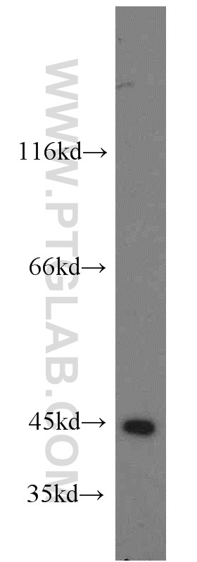
- Submitted by
- Proteintech Group (provider)
- Main image

- Experimental details
- HEK-293 cells were subjected to SDS PAGE followed by western blot with 20180-1-AP(PSAT1 antibody) at dilution of 1:500
- Sample type
- cell line
Supportive validation
- Submitted by
- Proteintech Group (provider)
- Main image

- Experimental details
- Immunofluorescent analysis of HepG2 cells, using PSAT1 antibody 20180-1-AP at 1:25 dilution and Rhodamine-labeled goat anti-rabbit IgG (red).
- Sample type
- cell line

Supportive validation
- Submitted by
- Proteintech Group (provider)
- Main image
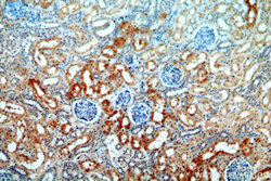
- Experimental details
- Immunohistochemical of paraffin-embedded human kidney using 20180-1-AP(PSAT1 antibody) at dilution of 1:100 (under 10x lens)
- Sample type
- tissue
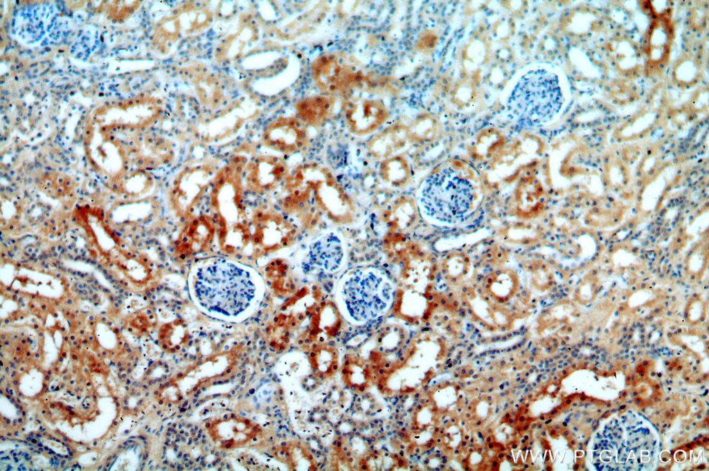
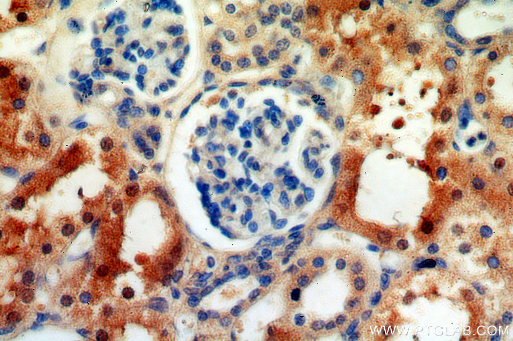
- Submitted by
- Proteintech Group (provider)
- Main image

- Experimental details
- Immunohistochemical of paraffin-embedded human kidney using 20180-1-AP(PSAT1 antibody) at dilution of 1:100 (under 40x lens)
- Sample type
- tissue