Explore
 Validate
 Learn
Learn Western blot
 Other assay
Antibody data
- Antibody Data
- Antigen structure
- References [1]
- Comments [0]
- Validations
- Other assay [1]
Submit
Validation data
Reference
Comment
Report error
- Product number
- PA5-30016 - Provider product page

- Provider
- Invitrogen Antibodies
- Product name
- EML1 Polyclonal Antibody
- Antibody type
- Polyclonal
- Antigen
- Recombinant full-length protein
- Description
- Recommended positive controls: A549, HeLa, HepG2, mouse brain. Predicted reactivity: Mouse (93%), Rat (93%). Store product as a concentrated solution. Centrifuge briefly prior to opening the vial.
- Reactivity
- Human, Mouse
- Host
- Rabbit
- Isotype
- IgG
- Vial size
- 100 μL
- Concentration
- 0.62 mg/mL
- Storage
- Store at 4°C short term. For long term storage, store at -20°C, avoiding freeze/thaw cycles.
Submitted references EML1 is essential for retinal photoreceptor migration and survival.
Poria D, Sun C, Santeford A, Kielar M, Apte RS, Kisselev OG, Chen S, Kefalov VJ
Scientific reports 2022 Feb 21;12(1):2897
Scientific reports 2022 Feb 21;12(1):2897
No comments: Submit comment
Supportive validation
- Submitted by
- Invitrogen Antibodies (provider)
- Main image
- Experimental details
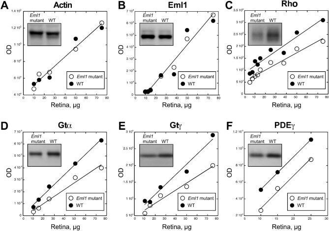
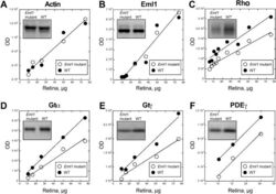
- Figure 6 Western blot analysis of Eml1 mutant retinas. Graphs showing optical density of Western blot bands against amount of total retina protein, n = 3. Eml1 mutant retinas (empty circles) as compared to the wild type retinas (filled circles). Linearity of plots demonstrates sub-saturating ECL signal ensuring direct quantitative comparison. ( A ) Actin, ( B ) EML1, ( C ) Rhodopsin, Rh, ( D ) transducin alpha, Gtalpha, ( E ) transducin gamma, Gtgamma, and ( F ) phosphodiesterase gamma (PDEgamma). Representative staining for each protein is shown in insets (for complete blot pictures see supplementary Fig. 4 ).