Explore
 Validate
 Learn
LearnMA5-24844
antibody from Invitrogen Antibodies
Targeting: TSPO
BZRP, DBI, IBP, MBR, mDRC, PBR, pk18, PKBS
 Western blot
 Immunocytochemistry
Antibody data
- Antibody Data
- Antigen structure
- References [2]
- Comments [0]
- Validations
- Immunocytochemistry [8]
- Flow cytometry [1]
- Other assay [5]
Submit
Validation data
Reference
Comment
Report error
- Product number
- MA5-24844 - Provider product page

- Provider
- Invitrogen Antibodies
- Product name
- TSPO Recombinant Rabbit Monoclonal Antibody (4H2)
- Antibody type
- Monoclonal
- Antigen
- Recombinant full-length protein
- Description
- Recombinant rabbit monoclonal antibodies are produced using in vitro expression systems. The expression systems are developed by cloning in the specific antibody DNA sequences from immunoreactive rabbits. Then, individual clones are screened to select the best candidates for production. The advantages of using recombinant rabbit monoclonal antibodies include: better specificity and sensitivity, lot-to-lot consistency, animal origin-free formulations, and broader immunoreactivity to diverse targets due to larger rabbit immune repertoire.
- Reactivity
- Human, Mouse
- Host
- Rabbit
- Isotype
- IgG
- Antibody clone number
- 4H2
- Vial size
- 100 μL
- Concentration
- 1 mg/mL
- Storage
- Store at 4°C short term. For long term storage, store at -20°C, avoiding freeze/thaw cycles.
Submitted references Osteopontin/secreted phosphoprotein-1 behaves as a molecular brake regulating the neuroinflammatory response to chronic viral infection.
Concentration, distribution, and influence of aging on the 18 kDa translocator protein in human brain: Implications for brain imaging studies.
Mahmud FJ, Du Y, Greif E, Boucher T, Dannals RF, Mathews WB, Pomper MG, Sysa-Shah P, Metcalf Pate KA, Lyons C, Carlson B, Chacona M, Brown AM
Journal of neuroinflammation 2020 Sep 17;17(1):273
Journal of neuroinflammation 2020 Sep 17;17(1):273
Concentration, distribution, and influence of aging on the 18 kDa translocator protein in human brain: Implications for brain imaging studies.
Tong J, Williams B, Rusjan PM, Mizrahi R, Lacapère JJ, McCluskey T, Furukawa Y, Guttman M, Ang LC, Boileau I, Meyer JH, Kish SJ
Journal of cerebral blood flow and metabolism : official journal of the International Society of Cerebral Blood Flow and Metabolism 2020 May;40(5):1061-1076
Journal of cerebral blood flow and metabolism : official journal of the International Society of Cerebral Blood Flow and Metabolism 2020 May;40(5):1061-1076
No comments: Submit comment
Supportive validation
- Submitted by
- Invitrogen Antibodies (provider)
- Main image
- Experimental details
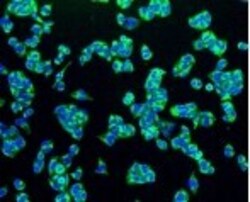
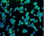
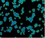
- Immunofluorescence analysis of TSPO in SW480 cells (green). Samples were treated with paraformaldehyde, permeabilized with 0.25% Triton and PBS and incubated with TSPO monoclonal antibody (Product # MA5-24844).

- Submitted by
- Invitrogen Antibodies (provider)
- Main image
- Experimental details
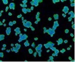
- Immunofluorescence analysis of TSPO in PC-3M cells (green). Samples were treated with paraformaldehyde, permeabilized with 0.25% Triton and PBS, and incubated with TSPO monoclonal antibody (Product # MA5-24844).

- Submitted by
- Invitrogen Antibodies (provider)
- Main image
- Experimental details
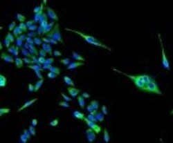
- Immunofluorescence analysis of TSPO in HeLa cells (green). Samples were treated with paraformaldehyde, permeabilized with 0.25% Triton and PBS, and incubated with TSPO monoclonal antibody (Product # MA5-24844).
- Submitted by
- Invitrogen Antibodies (provider)
- Main image
- Experimental details
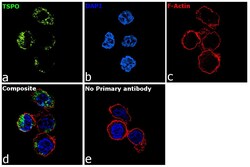
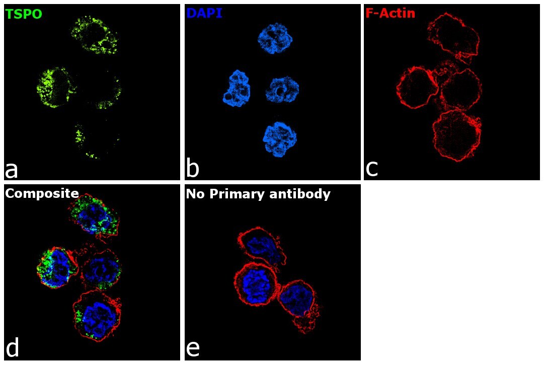
- Immunofluorescence analysis of TSPO was performed using THP-1 cells. The cells were fixed with 4% paraformaldehyde for 10 minutes, permeabilized with 0.1% Triton™ X-100 for 15 minutes, and blocked with 2% BSA for 1 hour at room temperature. The cells were labeled with TSPO Recombinant Rabbit Monoclonal Antibody (Product # MA5-24844) at 1:200 dilution in 0.1% BSA and incubated overnight at 4 degree and then labeled with Goat anti-Rabbit IgG (H+L) Superclonal™ Recombinant Secondary Antibody, Alexa Fluor® 488 (Product # A27034, 1:2000 dilution) for 45 minutes at room temperature (Panel a: green) in THP1 cells. Nuclei (Panel b: blue) were stained with ProLong™ Diamond Antifade Mountant with DAPI (Product # P36962). F-actin (Panel c: red) was stained with Rhodamine Phalloidin (Product # R415, 1:300). Panel d represents the merged image of HeLa cells showing nuclear membrane and cytosol localization for TSPO. Panel e represents control cells with no primary antibody to assess background. The images were captured at 60X magnification.
- Submitted by
- Invitrogen Antibodies (provider)
- Main image

- Experimental details
- Immunofluorescence analysis of TSPO in SW480 cells (green). Samples were treated with paraformaldehyde, permeabilized with 0.25% Triton and PBS and incubated with TSPO monoclonal antibody (Product # MA5-24844).
- Submitted by
- Invitrogen Antibodies (provider)
- Main image

- Experimental details
- Immunofluorescence analysis of TSPO in PC-3M cells (green). Samples were treated with paraformaldehyde, permeabilized with 0.25% Triton and PBS, and incubated with TSPO monoclonal antibody (Product # MA5-24844).

- Submitted by
- Invitrogen Antibodies (provider)
- Main image

- Experimental details
- Immunofluorescence analysis of TSPO in HeLa cells (green). Samples were treated with paraformaldehyde, permeabilized with 0.25% Triton and PBS, and incubated with TSPO monoclonal antibody (Product # MA5-24844).

- Submitted by
- Invitrogen Antibodies (provider)
- Main image

- Experimental details
- Immunofluorescence analysis of TSPO was performed using THP-1 cells. The cells were fixed with 4% paraformaldehyde for 10 minutes, permeabilized with 0.1% Triton™ X-100 for 15 minutes, and blocked with 2% BSA for 1 hour at room temperature. The cells were labeled with TSPO Recombinant Rabbit Monoclonal Antibody (Product # MA5-24844) at 1:200 dilution in 0.1% BSA and incubated overnight at 4 degree and then labeled with Goat anti-Rabbit IgG (Heavy Chain) Superclonal™ Recombinant Secondary Antibody, Alexa Fluor® 488 (Product # A27034, 1:2000 dilution) for 45 minutes at room temperature (Panel a: green) in THP1 cells. Nuclei (Panel b: blue) were stained with ProLong™ Diamond Antifade Mountant with DAPI (Product # P36962). F-actin (Panel c: red) was stained with Rhodamine Phalloidin (Product # R415, 1:300). Panel d represents the merged image of HeLa cells showing nuclear membrane and cytosol localization for TSPO. Panel e represents control cells with no primary antibody to assess background. The images were captured at 60X magnification.

Supportive validation
- Submitted by
- Invitrogen Antibodies (provider)
- Main image
- Experimental details
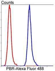
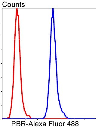
- Flow cytometry analysis of TSPO in HeLa cells (blue) and compared with an unlabeled control (cells without incubation with primary antibody; red). Samples were incubated with TSPO monoclonal antibody (Product # MA5-24844) at a dilution of 1:50 followed by Alexa Fluor 488-conjugated goat anti rabbit IgG.
Supportive validation
- Submitted by
- Invitrogen Antibodies (provider)
- Main image
- Experimental details
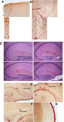
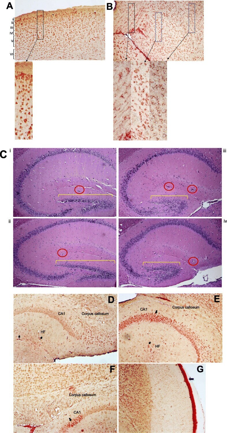
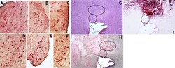
- Fig. 3 TSPO-Iba-1 double label immunoreactivity is abundant in cortical and hippocampal regions. a - f Representative samples of double-label immunostained 5 mum brain sections for TSPO (red) and Iba-1 (brown). a Multiple layers (i-vi) of the cortex can be seen from buffer-OPN + group. b Midbrain region from HIV-OPN + group, showing a diversity of microglia morphologies. c HE-stained hippocampal brain regions (i) Buffer-OPN + , (ii) Buffer-OPN - , (iii) HIV OPN + , (iv) HIV OPN - . Yellow bars, highlight the dentate gyrus and red circles indicate hippocampal fissures. d , e Hippocampus of HIV-OPN - . f Hippocampus in buffer-OPN - . g Meningeal inflammation in HIV OPN +
- Submitted by
- Invitrogen Antibodies (provider)
- Main image

- Experimental details
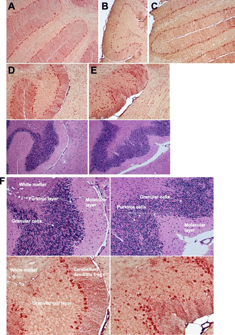
- Fig. 4 Robust TSPO immunoreactivity in a subset of neurons in the cerebellum. a-e Representative samples of double-label immunostained 5 mum brain sections for TSPO (red) and Iba-1 (brown) and their corresponding hematoxylin/eosin near adjacent section are shown a Buffer-OPN + , b HIV-OPN + , c HIV-OPN + , d Buffer-OPN - , and e HIV-OPN - . An ordered array of Purkinje neurons was not visible in HIV-infected mice lacking osteopontin ( e , HIV-OPN - ) compared to d HIV-OPN + . f Enlarged area from d , e is shown with the white matter, Purkinje, granular, and molecular layers of the cerebellum indicated by the arrows
- Submitted by
- Invitrogen Antibodies (provider)
- Main image
- Experimental details
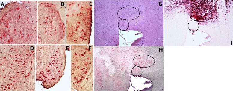
- Fig. 5 Strong TSPO immunoreactivity in a subset of tyrosine hydroxylase+ neurons in the striatum. a-f Representative samples of double-label immunostained 5 mum brain sections for TSPO (red) and Iba-1 (brown). a Buffer OPN+, b HIV OPN+, c HIV OPN+, d Buffer OPN-, e HIV OPN-, f Buffer OPN-. g hematoxylin/eosin stained section, and serial adjacent sections stained with h TSPO and i tyrosine hydroxylase. The black ovals demarcate the region of interest in each section
- Submitted by
- Invitrogen Antibodies (provider)
- Main image
- Experimental details
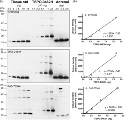
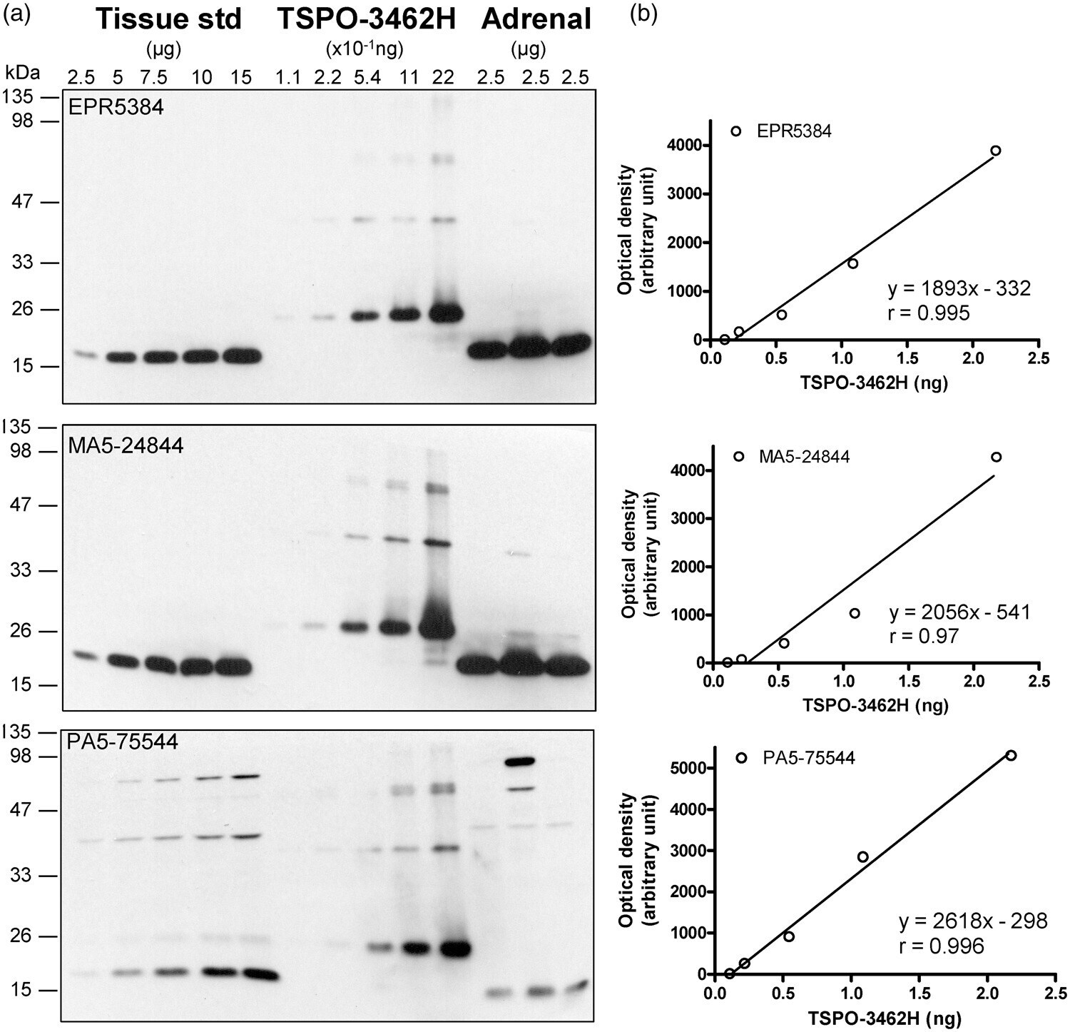
- Figure 1. Quantification of TSPO protein in autopsied human brain. (a) Immunoblots of TSPO in the pooled tissue standards, in the commercial N-His-tagged recombinant human TSPO-3462H and in human adrenal samples with monoclonal (EPR5384, 1:100K and MA5-24844, 1:30K dilution) and polyclonal (PA5-75544, 1:10K) antibodies. (b) Standard curves for the recombinant TSPO. Note more abundant TSPO in adrenal than in brain for both EPR5384 and MA5-24844, polymer protein bands of the recombinant TSPO, high molecular weight non-specific reactions for PA5-75544, and detection of a TSPO fragment but not 18 kDa TSPO by PA5-75544 in human adrenal samples.
- Submitted by
- Invitrogen Antibodies (provider)
- Main image
- Experimental details
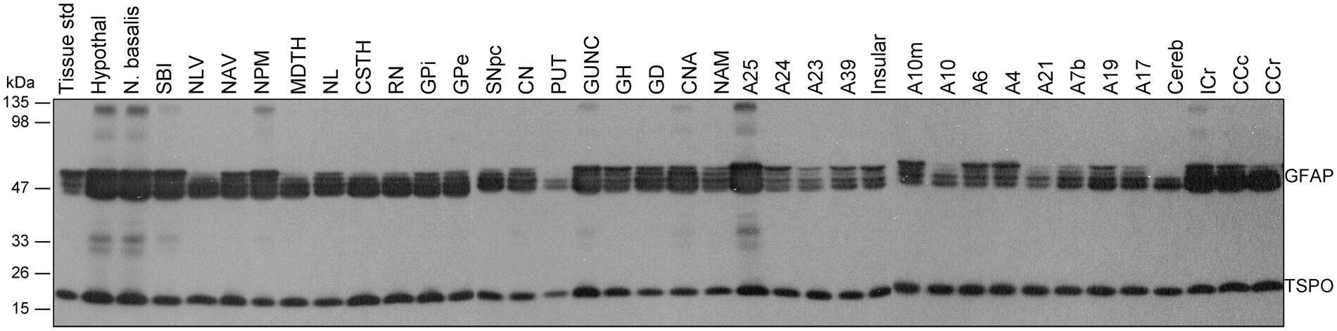
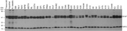
- Figure 2. Representative immunoblots of the regional distribution of TSPO and GFAP in autopsied human brain. A23: cingulate gyrus posterior; A24: cingulate gyrus anterior; A25: paraolfactory/subgenual gyrus; CCc: corpus callosum caudal; CCr: corpus callosum rostral; cereb: cerebellar cortex; CN: caudate; CNA: hippocampal Ammon's horn; CSTH: subthalamic nucleus; GD: dentate gyrus; GH: hippocampal gyrus; GPe: globus pallidus external; GPi: globus pallidus internal; GUNC: gyrus of uncus; hypothal: hypothalamus; ICr: internal capsule rostral; MDTH: mediodorsal thalamus; NAM: amygdala; NAV: anterior ventral nucleus of thalamus; N. basalis: nucleus basalis; NL: nucleus lateralis of thalamus; NLV: lateral ventral nucleus of thalamus; NPM: medial pulvinar of thalamus; PUT: putamen; RN: red nucleus; SBI: substantia innominata; SNpc: substantia nigra pars compacta.