 Explore
 Validate
 Learn
LearnPA5-67938
antibody from Invitrogen Antibodies
Targeting: TSPO
BZRP, DBI, IBP, MBR, mDRC, PBR, pk18, PKBS
 Western blot
Antibody data
- Antibody Data
- Antigen structure
- References [0]
- Comments [0]
- Validations
- Western blot [2]
- Immunocytochemistry [1]
Submit
Validation data
Reference
Comment
Report error
- Product number
- PA5-67938 - Provider product page

- Provider
- Invitrogen Antibodies
- Product name
- TSPO Polyclonal Antibody
- Antibody type
- Polyclonal
- Antigen
- Synthetic peptide
- Reactivity
- Human
- Host
- Rabbit
- Isotype
- IgG
- Vial size
- 100 µL
- Concentration
- 1 mg/mL
- Storage
- -20°C
No comments: Submit comment
Supportive validation
- Submitted by
- Invitrogen Antibodies (provider)
- Main image

- Experimental details
- Western blot analysis of TSPO in Jurkat whole cell lysates using a TSPO Polyclonal Antibody (Product # PA5-67938).
- Submitted by
- Invitrogen Antibodies (provider)
- Main image

- Experimental details
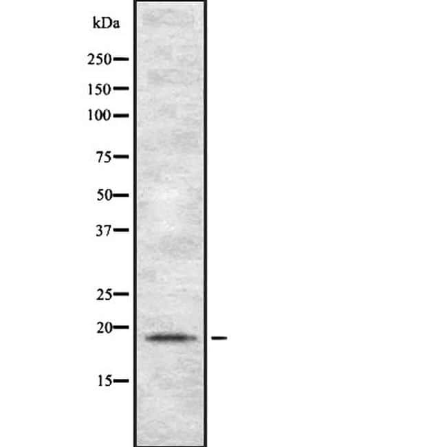
- Western blot was performed using TSPO Recombinant Rabbit Monoclonal Antibody (Product # PA5-67938) and a 18 kDa band corresponding to TSPO was observed across cell lines tested and increased upon TPA treatment in THP-1; the expression was low to nil in SK-OV-3, HeLa and Hep G2. Membrane enriched extracts (30 µg lysate) of THP-1 treated with TPA (50 ng/mL for 24 Hours) (Lane 1), THP-1 (Lane 2), Raw 264.7 (Lane 3), SK-O-V3 (Lane 4), HeLa (Lane 5) and Hep G2 (Lane 6) were electrophoresed using Novex® NuPAGE® 16 % Tricine gel (Product # EC6695BOX). Resolved proteins were then transferred onto a nitrocellulose membrane (Product # IB23001) by iBlot® 2 Dry Blotting System (Product # IB21001). The blot was probed with the primary antibody (1:1000 dilution) and detected by chemiluminescence with Goat anti-Rabbit IgG (H+L), Superclonal™ Recombinant Secondary Antibody, HRP (Product # A27036, 1:4000 dilution) using the iBright FL 1000 (Product # A32752). Chemiluminescent detection was performed using Novex® ECL Chemiluminescent Substrate Reagent Kit (Product # WP20005).

Supportive validation
- Submitted by
- Invitrogen Antibodies (provider)
- Main image
- Experimental details
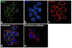
- Immunofluorescence analysis of TSPO was performed using THP-1 cells. The cells were fixed with 4% paraformaldehyde for 10 minutes, permeabilized with 0.1% Triton™ X-100 for 15 minutes, and blocked with 2% BSA for 1 hour at room temperature. The cells were labeled with TSPO Recombinant Rabbit Monoclonal Antibody (Product # PA5-67938) at 1:200 dilution in 0.1% BSA and incubated overnight at 4 degree and then labeled with Goat anti-Rabbit IgG (H+L) Superclonal™ Recombinant Secondary Antibody, Alexa Fluor® 488 (Product # A27034, 1:2000 dilution) for 45 minutes at room temperature (Panel a: green) in HeLa cells. Nuclei (Panel b: blue) were stained with ProLong™ Diamond Antifade Mountant with DAPI (Product # P36962). F-actin (Panel c: red) was stained with Rhodamine Phalloidin (Product # R415, 1:300). Panel d represents the merged image of THP-1 cells showing nuclear membrane and cytosol localization for TSPO. Panel e represents control cells with no primary antibody to assess background. The images were captured at 60X magnification.
