 Explore
 Validate
 Learn
LearnPA5-75544
antibody from Invitrogen Antibodies
Targeting: TSPO
BZRP, DBI, IBP, MBR, mDRC, PBR, pk18, PKBS
 Western blot
Antibody data
- Antibody Data
- Antigen structure
- References [4]
- Comments [0]
- Validations
- Western blot [2]
- Immunocytochemistry [1]
- Other assay [4]
Submit
Validation data
Reference
Comment
Report error
- Product number
- PA5-75544 - Provider product page

- Provider
- Invitrogen Antibodies
- Product name
- TSPO Polyclonal Antibody
- Antibody type
- Polyclonal
- Antigen
- Synthetic peptide
- Description
- The antibody was affinity-purified from rabbit antiserum by affinity-chromatography using epitope-specific immunogen and the purity is > 95% (by SDS-PAGE).
- Reactivity
- Human, Mouse, Rat
- Host
- Rabbit
- Isotype
- IgG
- Vial size
- 100 µL
- Concentration
- 1 mg/mL
- Storage
- Store at 4°C short term. For long term storage, store at -20°C, avoiding freeze/thaw cycles.
Submitted references Neurogenic Potential of the 18-kDa Mitochondrial Translocator Protein (TSPO) in Pluripotent P19 Stem Cells.
The peroxisome proliferator-activated receptor gamma (PPARγ) agonist, rosiglitazone, ameliorates neurofunctional and neuroinflammatory abnormalities in a rat model of Gulf War Illness.
Concentration, distribution, and influence of aging on the 18 kDa translocator protein in human brain: Implications for brain imaging studies.
TSPO ligands prevent the proliferation of vascular smooth muscle cells and attenuate neointima formation through AMPK activation.
González-Blanco L, Bermejo-Millo JC, Oliveira G, Potes Y, Antuña E, Menéndez-Valle I, Vega-Naredo I, Coto-Montes A, Caballero B
Cells 2021 Oct 17;10(10)
Cells 2021 Oct 17;10(10)
The peroxisome proliferator-activated receptor gamma (PPARγ) agonist, rosiglitazone, ameliorates neurofunctional and neuroinflammatory abnormalities in a rat model of Gulf War Illness.
Keledjian K, Tsymbalyuk O, Semick S, Moyer M, Negoita S, Kim K, Ivanova S, Gerzanich V, Simard JM
PloS one 2020;15(11):e0242427
PloS one 2020;15(11):e0242427
Concentration, distribution, and influence of aging on the 18 kDa translocator protein in human brain: Implications for brain imaging studies.
Tong J, Williams B, Rusjan PM, Mizrahi R, Lacapère JJ, McCluskey T, Furukawa Y, Guttman M, Ang LC, Boileau I, Meyer JH, Kish SJ
Journal of cerebral blood flow and metabolism : official journal of the International Society of Cerebral Blood Flow and Metabolism 2020 May;40(5):1061-1076
Journal of cerebral blood flow and metabolism : official journal of the International Society of Cerebral Blood Flow and Metabolism 2020 May;40(5):1061-1076
TSPO ligands prevent the proliferation of vascular smooth muscle cells and attenuate neointima formation through AMPK activation.
Wu LP, Gong ZF, Wang H, Zhou ZS, Zhang MM, Liu C, Ren HM, Yang J, Han Y, Zeng CY
Acta pharmacologica Sinica 2020 Jan;41(1):34-46
Acta pharmacologica Sinica 2020 Jan;41(1):34-46
No comments: Submit comment
Supportive validation
- Submitted by
- Invitrogen Antibodies (provider)
- Main image

- Experimental details
- Western blot analysis of TSPO in Lane 1: PMVEC whole cell lysate (40 µg), Lane 2: HepG2 whole cell lysate (40 µg), Lane 3: 3T3-L1 whole cell lysate (40 µg), Lane 4: A2780 whole cell lysate (40 µg), Lane 5: A549 whole cell lysate (40 µg). Samples were incubated with TSPO polyclonal antibody (Product # PA5-75544) at a dilution of 1:2000.

- Submitted by
- Invitrogen Antibodies (provider)
- Main image

- Experimental details
- Western blot was performed using TSPO Recombinant Rabbit Monoclonal Antibody (Product # PA5-75544) and a 18 kDa band corresponding to TSPO was observed across cell lines tested and increased upon TPA treatment in THP-1; the expression was low to nil in SK-OV-3, HeLa and Hep G2. Membrane enriched extracts (30 µg lysate) of THP-1 treated with TPA (50 ng/mL for 24 Hours) (Lane 1), THP-1 (Lane 2), Raw 264.7 (Lane 3), SK-O-V3 (Lane 4), HeLa (Lane 5) and HepG2 (Lane 6) were electrophoresed using Novex® NuPAGE® 16 % Tricine gel (Product # EC6695BOX). Resolved proteins were then transferred onto a nitrocellulose membrane (Product # IB23001) by iBlot® 2 Dry Blotting System (Product # IB21001). The blot was probed with the primary antibody (1:500 dilution) and detected by chemiluminescence with Goat anti-Rabbit IgG (H+L), Superclonal™ Recombinant Secondary Antibody, HRP (Product # A27036, 1:4000 dilution) using the iBright FL 1000 (Product # A32752). Chemiluminescent detection was performed using Novex® ECL Chemiluminescent Substrate Reagent Kit (Product # WP20005).

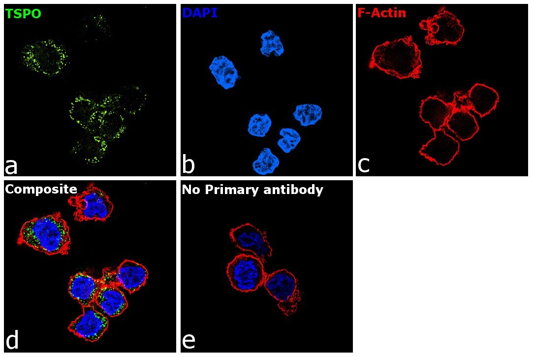
Supportive validation
- Submitted by
- Invitrogen Antibodies (provider)
- Main image

- Experimental details
- Immunofluorescence analysis of TSPO was performed using THP-1 cells. The cells were fixed with 4% paraformaldehyde for 10 minutes, permeabilized with 0.1% Triton™ X-100 for 15 minutes, and blocked with 2% BSA for 1 hour at room temperature. The cells were labeled with TSPO Recombinant Rabbit Monoclonal Antibody (Product # PA5-75544) at 1:200 dilution in 0.1% BSA and incubated overnight at 4 degree and then labeled with Goat anti-Rabbit IgG (H+L) Superclonal™ Recombinant Secondary Antibody, Alexa Fluor® 488 (Product # A27034, 1:2000 dilution) for 45 minutes at room temperature (Panel a: green) in HeLa cells. Nuclei (Panel b: blue) were stained with ProLong™ Diamond Antifade Mountant with DAPI (Product # P36962). F-actin (Panel c: red) was stained with Rhodamine Phalloidin (Product # R415, 1:300). Panel d represents the merged image of THP-1 cells showing nuclear membrane and cytosol localization for TSPO. Panel e represents control cells with no primary antibody to assess background. The images were captured at 60X magnification.
Supportive validation
- Submitted by
- Invitrogen Antibodies (provider)
- Main image

- Experimental details
- Fig 5 Astrocytosis following 33-day GWE. A,B: Immunolabeling for GFAP in hippocampus (HC) and lateral amygdala (LA) in naive (CTR), GWE-VEH and GWE-ROSI rats; nuclear staining with DAPI (blue) is also shown; bars , 100 mum. C: Quantification of GFAP in the two brain regions for the three groups, as indicated; 5 rats per group; **, P

- Submitted by
- Invitrogen Antibodies (provider)
- Main image

- Experimental details
- Figure 1 ( A ) Representative immunoblot of the TSPO protein levels in undifferentiated P19 stem cells (SC) and cells treated with 1 muM retinoid acid (RA) or 50 muM PK 11195 (PK 11195) during 4 days of treatment. ( B ). Bar chart shows quantification of the optical densities (OD) from three independent cell cultures and treatments. Data are expressed as means +- SEM. *** p < 0.001 versus SC. Protein shows a representative ponceau staining of one of the experiments. The complete ponceau staining for TSPO antibody is shown in the Supplementary Figure S1 . Although the entire membrane of ponceau S was used for total protein normalization, only a representative section of the membrane is displayed in this image. TSPO, the 18-kDa mitochondrial translocator protein.

- Submitted by
- Invitrogen Antibodies (provider)
- Main image

- Experimental details
- Figure 1. Quantification of TSPO protein in autopsied human brain. (a) Immunoblots of TSPO in the pooled tissue standards, in the commercial N-His-tagged recombinant human TSPO-3462H and in human adrenal samples with monoclonal (EPR5384, 1:100K and MA5-24844, 1:30K dilution) and polyclonal (PA5-75544, 1:10K) antibodies. (b) Standard curves for the recombinant TSPO. Note more abundant TSPO in adrenal than in brain for both EPR5384 and MA5-24844, polymer protein bands of the recombinant TSPO, high molecular weight non-specific reactions for PA5-75544, and detection of a TSPO fragment but not 18 kDa TSPO by PA5-75544 in human adrenal samples.

- Submitted by
- Invitrogen Antibodies (provider)
- Main image

- Experimental details
- Figure 2. Representative immunoblots of the regional distribution of TSPO and GFAP in autopsied human brain. A23: cingulate gyrus posterior; A24: cingulate gyrus anterior; A25: paraolfactory/subgenual gyrus; CCc: corpus callosum caudal; CCr: corpus callosum rostral; cereb: cerebellar cortex; CN: caudate; CNA: hippocampal Ammon's horn; CSTH: subthalamic nucleus; GD: dentate gyrus; GH: hippocampal gyrus; GPe: globus pallidus external; GPi: globus pallidus internal; GUNC: gyrus of uncus; hypothal: hypothalamus; ICr: internal capsule rostral; MDTH: mediodorsal thalamus; NAM: amygdala; NAV: anterior ventral nucleus of thalamus; N. basalis: nucleus basalis; NL: nucleus lateralis of thalamus; NLV: lateral ventral nucleus of thalamus; NPM: medial pulvinar of thalamus; PUT: putamen; RN: red nucleus; SBI: substantia innominata; SNpc: substantia nigra pars compacta.
