Explore
 Validate
 Learn
Learn Western blot
 Immunocytochemistry
Antibody data
- Antibody Data
- Antigen structure
- References [0]
- Comments [0]
- Validations
- Western blot [2]
- Immunocytochemistry [3]
- Immunoprecipitation [4]
- Immunohistochemistry [2]
Submit
Validation data
Reference
Comment
Report error
- Product number
- LS-C185381 - Provider product page

- Provider
- LSBio
- Product name
- HAUS8 Antibody (aa166-360) LS-C185381
- Antibody type
- Polyclonal
- Description
- Immunoaffinity purified
- Reactivity
- Human
- Host
- Rabbit
- Isotype
- IgG
- Storage
- Keep as concentrated solution. Aliquot and store at -20°C or below. Avoid multiple freeze-thaw cycles.
No comments: Submit comment
Enhanced validation
- Submitted by
- LSBio (provider)
- Enhanced method
- Genetic validation
- Main image
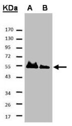
- Experimental details
- Sample. A: His-Hice1 (2x), B: His-Hice1 (1x). 7.5% SDS PAGE. HAUS8 antibody diluted at 1:500

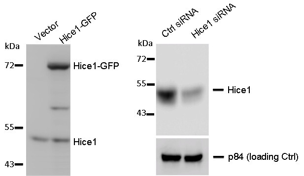
- Submitted by
- LSBio (provider)
- Enhanced method
- Genetic validation
- Main image
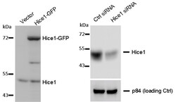
- Experimental details
- WB to detect cellular Hice1 and Hice1-GFP expressed in human osteosarcoma U2OS cells (left image) , and Hice1 upon siRNA treatment (right image) using HAUS8 antibody at 1:1000 dilution. Nuclear matrix protein p84 is a loading control, blotted with p84 antibody (clone 5E10) GTX70220 at 1:5000 dilution.
Supportive validation
- Submitted by
- LSBio (provider)
- Enhanced method
- Genetic validation
- Main image

- Experimental details
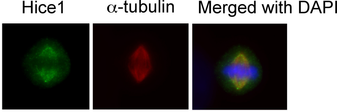
- Immunofluorescence of human osteosarcoma cell line U2OS using HAUS8 antibody at 1:50-1:200 dilution.
- Submitted by
- LSBio (provider)
- Main image
- Experimental details
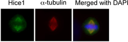
- Immunofluorescence of human osteosarcoma cell line U2OS using HAUS8 antibody at 1:50-1:200 dilution.

- Submitted by
- LSBio (provider)
- Main image

- Experimental details
- Immunofluorescence of human osteosarcoma cell line U2OS using HAUS8 antibody at 1:50-1:200 dilution.

Supportive validation
- Submitted by
- LSBio (provider)
- Enhanced method
- Genetic validation
- Main image

- Experimental details
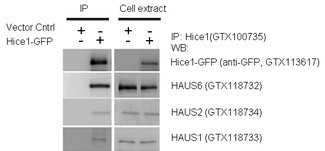
- IP-WB assay to show that Hice1 co-immunoprecipitated with other Augmin components HAUS6, HAUS2 and HAUS1 in U2OS cells.

- Submitted by
- LSBio (provider)
- Main image

- Experimental details
- IP-WB assay to show that Hice1 co-immunoprecipitated with other Augmin components HAUS6, HAUS2 and HAUS1 in U2OS cells.

- Submitted by
- LSBio (provider)
- Main image

- Experimental details
- IP-WB assay to show that Hice1 co-immunoprecipitated with other Augmin components HAUS6, HAUS2 and HAUS1 in U2OS cells.
- Submitted by
- LSBio (provider)
- Main image

- Experimental details
- IP-WB assay to show that Hice1 co-immunoprecipitated with other Augmin components HAUS6, HAUS2 and HAUS1 in U2OS cells.

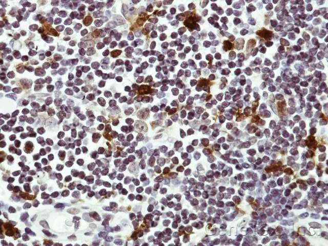
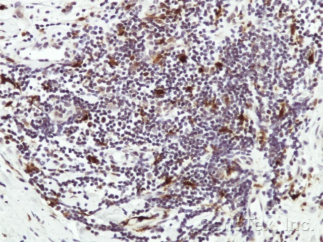
Supportive validation
- Submitted by
- LSBio (provider)
- Enhanced method
- Genetic validation
- Main image
- Experimental details
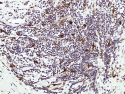
- IHC of paraffin-embedded Human lymph tissue using HAUS8 antibody at 1:50 dilution.
- Submitted by
- LSBio (provider)
- Enhanced method
- Genetic validation
- Main image

- Experimental details
- IHC of paraffin-embedded Human lymph tissue using HAUS8 antibody at 1:50 dilution.