Explore
 Validate
 Learn
LearnRP1056
antibody from Boster Biological Technology
Targeting: NCOA1
bHLHe74, F-SRC-1, KAT13A, NCoA-1, RIP160, SRC1
 Western blot
 Immunocytochemistry
Antibody data
- Antibody Data
- Antigen structure
- References [1]
- Comments [0]
- Validations
- Western blot [1]
Submit
Validation data
Reference
Comment
Report error
- Product number
- RP1056 - Provider product page

- Provider
- Boster Biological Technology
- Product name
- Anti-KAT13A/SRC1/NCOA1 Antibody
- Antibody type
- Polyclonal
- Description
- Polyclonal antibody for SRC 1/NCOA1 detection. Host: Rabbit.Size: 100μg/vial. Tested applications: WB, ICC/IF, FCM. Reactive species: Human. SRC 1/NCOA1 information: Molecular Weight: 156757 MW; Subcellular Localization: Nucleus ; Tissue Specificity: Widely expressed.
- Reactivity
- Human
- Host
- Rabbit
- Vial size
- 100μg/vial
- Concentration
- Add 0.2ml of distilled water will yield a concentration of 500ug/ml.
- Storage
- At -20°C for one year. After reconstitution, at 4°C for one month. It can also be aliquoted and stored frozen at -20°C for a longer time. Avoid repeated freezing and thawing.
- Handling
- Add 0.2ml of distilled water will yield a concentration of 500ug/ml.
Submitted references Explore the mechanism and substance basis of Mahuang FuziXixin Decoction for the treatment of lung cancer based on network pharmacology and molecular docking.
Zhang W, Tian W, Wang Y, Jin X, Guo H, Wang Y, Tang Y, Yao X
Computers in biology and medicine 2022 Dec;151(Pt A):106293
Computers in biology and medicine 2022 Dec;151(Pt A):106293
No comments: Submit comment
Supportive validation
- Submitted by
- Boster Biological Technology (provider)
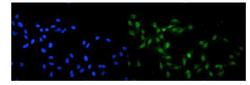
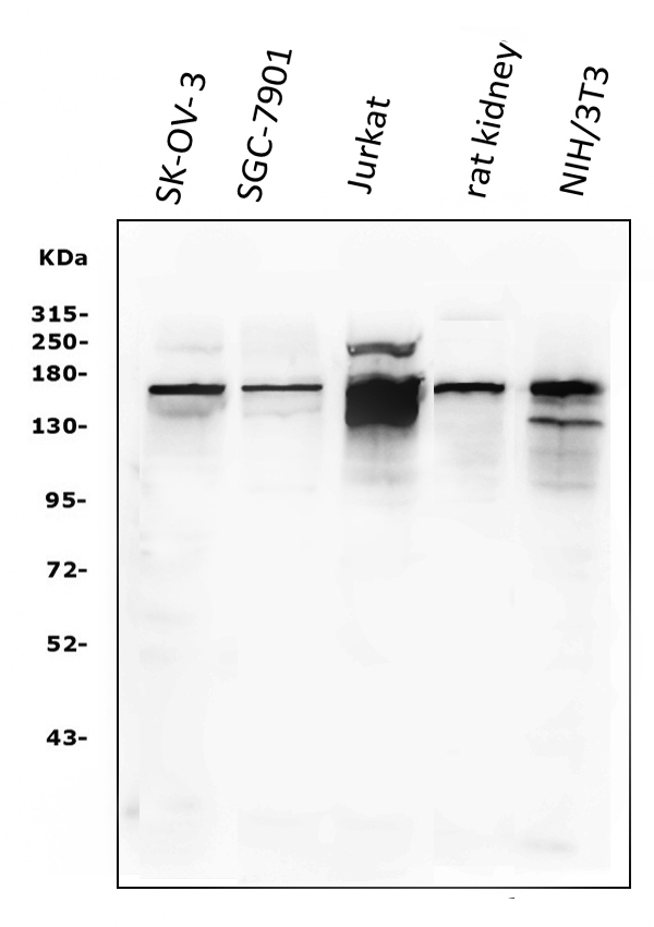
- Main image

- Experimental details
- Western blot analysis of KAT13A/SRC1 using anti-KAT13A/SRC1 antibody (RP1056). Electrophoresis was performed on a 5-20% SDS-PAGE gel at 70V (Stacking gel) / 90V (Resolving gel) for 2-3 hours. The sample well of each lane was loaded with 50ug of sample under reducing conditions. Lane 1: SK-OV-3 whole cell lysates, Lane 2: SGC-7901 whole cell lysates, Lane 3: Jurkat whole cell lysates, Lane 4: rat kidney tissue lysates, Lane 5: NIH/3T3 whole cell lysates. After Electrophoresis, proteins were transferred to a Nitrocellulose membrane at 150mA for 50-90 minutes. Blocked the membrane with 5% Non-fat Milk/ TBS for 1.5 hour at RT. The membrane was incubated with rabbit anti-KAT13A/SRC1 antigen affinity purified polyclonal antibody (Catalog # RP1056) at 0.5 μg/mL overnight at 4°C, then washed with TBS-0.1%Tween 3 times with 5 minutes each and probed with a goat anti-rabbit IgG-HRP secondary antibody at a dilution of 1:5000 for 1.5 hour at RT. The signal is developed using an Enhanced Chemiluminescent detection (ECL) kit (Catalog # EK1002) with Tanon 5200 system. A specific band was detected for KAT13A/SRC1 at approximately 157KD. The expected band size for KAT13A/SRC1 is at 157KD.
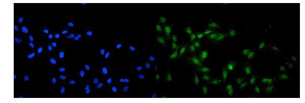
- Additional image