Explore
 Validate
 Learn
LearnHPA034537
antibody from Atlas Antibodies
Targeting: SAMM50
CGI-51, OMP85, SAM50, TOB55, TRG-3, YNL026W
 Western blot
 Immunocytochemistry
Antibody data
- Antibody Data
- Antigen structure
- References [4]
- Comments [0]
- Validations
- Immunocytochemistry [1]
- Immunohistochemistry [1]
Submit
Validation data
Reference
Comment
Report error
- Product number
- HPA034537 - Provider product page

- Provider
- Atlas Antibodies
- Proper citation
- Atlas Antibodies Cat#HPA034537, RRID:AB_10602794
- Product name
- Anti-SAMM50
- Antibody type
- Polyclonal
- Description
- Polyclonal Antibody against Human SAMM50, Gene description: SAMM50 sorting and assembly machinery component, Alternative Gene Names: CGI-51, OMP85, TOB55, TRG-3, YNL026W, Validated applications: ICC, IHC, WB, Uniprot ID: Q9Y512, Storage: Store at +4°C for short term storage. Long time storage is recommended at -20°C.
- Reactivity
- Human, Mouse, Rat
- Host
- Rabbit
- Conjugate
- Unconjugated
- Isotype
- IgG
- Vial size
- 100 µl
- Concentration
- 0.3 mg/ml
- Storage
- Store at +4°C for short term storage. Long time storage is recommended at -20°C.
- Handling
- The antibody solution should be gently mixed before use.
Submitted references Compositionally unique mitochondria in filopodia support cellular migration.
CARD19 Interacts with Mitochondrial Contact Site and Cristae Organizing System Constituent Proteins and Regulates Cristae Morphology.
Identification and characterization of protein N-myristoylation occurring on four human mitochondrial proteins, SAMM50, TOMM40, MIC19, and MIC25
Identification of Human N-Myristoylated Proteins from Human Complementary DNA Resources by Cell-Free and Cellular Metabolic Labeling Analyses
Marlar-Pavey M, Tapias-Gomez D, Mettlen M, Friedman JR
bioRxiv : the preprint server for biology 2024 Jun 21;
bioRxiv : the preprint server for biology 2024 Jun 21;
CARD19 Interacts with Mitochondrial Contact Site and Cristae Organizing System Constituent Proteins and Regulates Cristae Morphology.
Rios KE, Zhou M, Lott NM, Beauregard CR, McDaniel DP, Conrads TP, Schaefer BC
Cells 2022 Mar 31;11(7)
Cells 2022 Mar 31;11(7)
Identification and characterization of protein N-myristoylation occurring on four human mitochondrial proteins, SAMM50, TOMM40, MIC19, and MIC25
Saad J, Utsumi T, Matsuzaki K, Kiwado A, Tanikawa A, Kikkawa Y, Hosokawa T, Otsuka A, Iuchi Y, Kobuchi H, Moriya K
PLOS ONE 2018;13(11):e0206355
PLOS ONE 2018;13(11):e0206355
Identification of Human N-Myristoylated Proteins from Human Complementary DNA Resources by Cell-Free and Cellular Metabolic Labeling Analyses
Saad J, Takamitsu E, Otsuka M, Haebara T, Yano M, Matsuzaki K, Kobuchi H, Moriya K, Utsumi T
PLOS ONE 2015;10(8):e0136360
PLOS ONE 2015;10(8):e0136360
No comments: Submit comment
Supportive validation
- Submitted by
- Atlas Antibodies (provider)
- Main image

- Experimental details
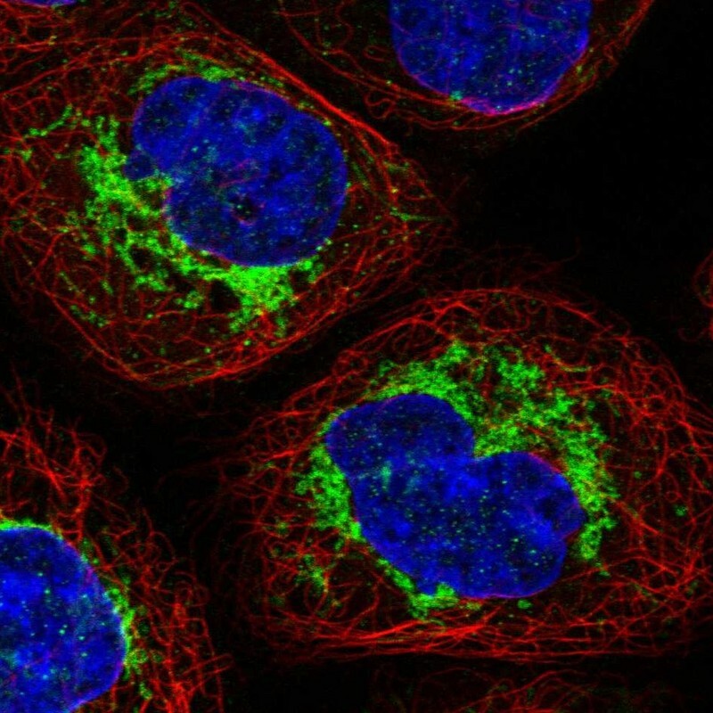
- Immunofluorescent staining of human cell line A-431 shows localization to mitochondria.
- Sample type
- Human
Supportive validation
- Submitted by
- Atlas Antibodies (provider)
- Enhanced method
- Orthogonal validation
- Main image
- Experimental details
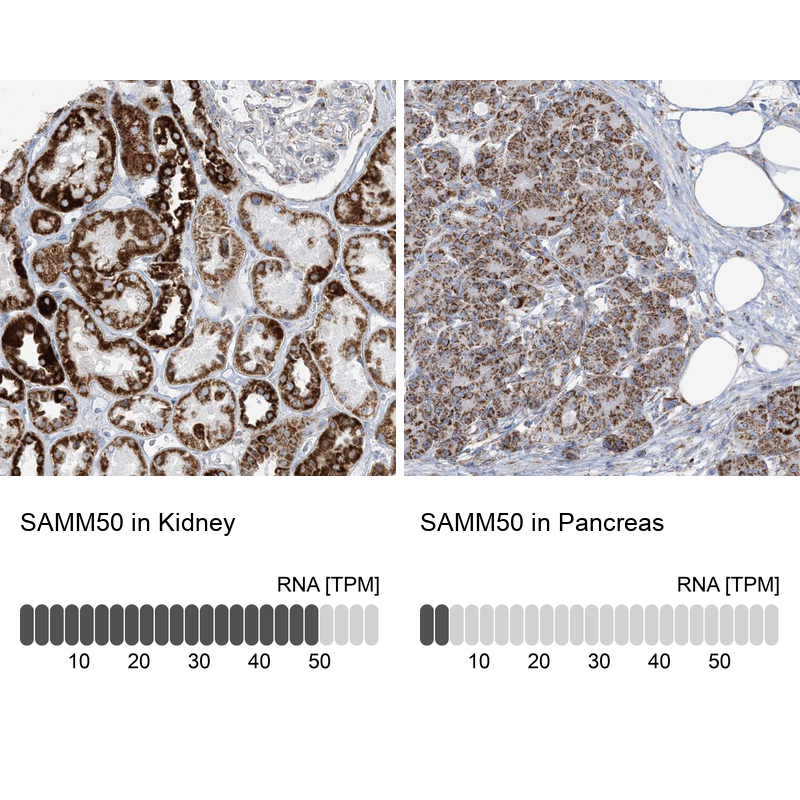
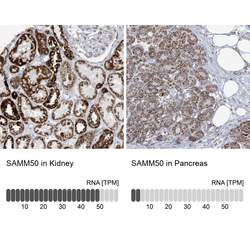
- Immunohistochemistry analysis in human kidney and pancreas tissues using HPA034537 antibody. Corresponding SAMM50 RNA-seq data are presented for the same tissues.
- Sample type
- Human
- Protocol
- Protocol