Explore
 Validate
 Learn
Learn Western blot
 ELISA
ELISA Immunocytochemistry
Antibody data
- Antibody Data
- Antigen structure
- References [0]
- Comments [0]
- Validations
- Western blot [2]
- Immunohistochemistry [1]
- Flow cytometry [1]
Submit
Validation data
Reference
Comment
Report error
- Product number
- AP53171PU-N - Provider product page

- Provider
- Acris Antibodies GmbH
- Proper citation
- Acris Antibodies GmbH Cat#AP53171PU-N, RRID:AB_11146385
- Product name
- anti PARVB (N-term)
- Antibody type
- Polyclonal
- Antigen
- KLH conjugated synthetic peptide between 69-99 amino acids from the N-terminal region of human PARVB
- Reactivity
- Human, Mouse
- Host
- Rabbit
- Vial size
- 0.1 mg
- Concentration
- 0.25 mg/ml
No comments: Submit comment
Supportive validation
- Submitted by
- Acris Antibodies GmbH (provider)
- Main image
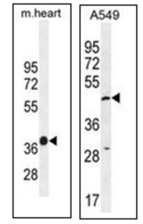
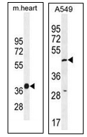
- Experimental details
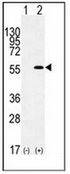
- Western blot analysis of PARVB Antibody (N-term) Cat.-No AP53171PU-N in A549 cell line lysates and in mouse heart tissue lysates (35ug/lane). This demonstrates the PARVB antibody detected the PARVB protein (arrow).
- Submitted by
- Acris Antibodies GmbH (provider)
- Main image
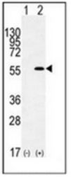
- Experimental details
- Western blot analysis of PARVB (arrow) using PARVB Antibody (N-term) Cat.-No AP53171PU-N. 293 cell lysates (2 ug/lane) either nontransfected (Lane 1) or transiently transfected (Lane 2) with the PARVB gene.
Supportive validation
- Submitted by
- Acris Antibodies GmbH (provider)
- Main image
- Experimental details
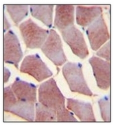
- Immunohistochemistry analysis in formalin fixed and paraffin embedded human skeletal muscle reacted with PARVB Antibody (N-term) Cat.-No AP53171PU-N, which was peroxidase conjugated to the secondary antibody and followed by DAB staining. This data demonstrates the use of PARVB Antibody (N-term) for immunohistochemistry. Clinical relevance has not been evaluated.

Supportive validation
- Submitted by
- Acris Antibodies GmbH (provider)
- Main image

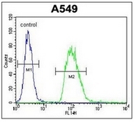
- Experimental details
- Flow cytometric analysis of A549 cells using PARVB Antibody (N-term) Cat.-No AP53171PU-N (right histogram) compared to a negative control cell (left histogram). FITC-conjugated goat-anti-rabbit secondary antibodies were used for the analysis.