Explore
 Validate
 Learn
Learn Western blot
 Immunocytochemistry
Antibody data
- Antibody Data
- Antigen structure
- References [3]
- Comments [0]
- Validations
- Immunocytochemistry [2]
- Immunohistochemistry [5]
- Other assay [1]
Submit
Validation data
Reference
Comment
Report error
- Product number
- PA5-29653 - Provider product page

- Provider
- Invitrogen Antibodies
- Product name
- ChAT Polyclonal Antibody
- Antibody type
- Polyclonal
- Antigen
- Recombinant full-length protein
- Description
- Recommended positive controls: IMR32, mouse brain, rat brain, DDDDK-tagged CHAT-transfected 293T. Predicted reactivity: Mouse (93%), Rat (92%), Pig (94%). Store product as a concentrated solution. Centrifuge briefly prior to opening the vial.
- Reactivity
- Human, Mouse, Rat
- Host
- Rabbit
- Isotype
- IgG
- Vial size
- 100 μL
- Concentration
- 0.15 mg/mL
- Storage
- Store at 4°C short term. For long term storage, store at -20°C, avoiding freeze/thaw cycles.
Submitted references Focal impaired awareness seizures in a rodent model: A functional anatomy.
Targeting the tamoxifen receptor within sodium channels to block osteoarthritic pain.
Angiopoietin-1 Knockout Mice as a Genetic Model of Open-Angle Glaucoma.
Adotevi N, Kapur J
Epilepsia open 2022 Mar;7(1):110-123
Epilepsia open 2022 Mar;7(1):110-123
Targeting the tamoxifen receptor within sodium channels to block osteoarthritic pain.
McCollum MM, Larmore M, Ishihara S, Ng LCT, Kimura LF, Guadarrama E, Ta MC, Vien TN, Frost GB, Scheidt KA, Miller RE, DeCaen PG
Cell reports 2022 Aug 23;40(8):111248
Cell reports 2022 Aug 23;40(8):111248
Angiopoietin-1 Knockout Mice as a Genetic Model of Open-Angle Glaucoma.
Thomson BR, Grannonico M, Liu F, Liu M, Mendapara P, Xu Y, Liu X, Quaggin SE
Translational vision science & technology 2020 Mar;9(4):16
Translational vision science & technology 2020 Mar;9(4):16
No comments: Submit comment
Supportive validation
- Submitted by
- Invitrogen Antibodies (provider)
- Main image

- Experimental details
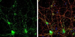
- Immunocytochemistry-Immunofluorescence analysis of ChAT was performed in DIV14 rat E18 primary cortical neurons fixed in 4% paraformaldehyde at RT for 15 min. Green: ChAT Polyclonal Antibody (Product # PA5-29653) diluted at 1:500. Red: beta Tubulin 3/ Tuj1, stained by beta Tubulin 3/ Tuj1 antibody. Blue: Fluoroshield with DAPI.

- Submitted by
- Invitrogen Antibodies (provider)
- Main image
- Experimental details
- Immunocytochemistry-Immunofluorescence analysis of ChAT was performed in DIV14 rat E18 primary cortical neurons fixed in 4% paraformaldehyde at RT for 15 min. Green: ChAT Polyclonal Antibody (Product # PA5-29653) diluted at 1:500. Red: beta Tubulin 3/ Tuj1, stained by beta Tubulin 3/ Tuj1 antibody. Blue: Fluoroshield with DAPI.

Supportive validation
- Submitted by
- Invitrogen Antibodies (provider)
- Main image

- Experimental details
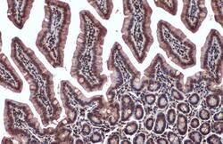
- Immunohistochemistry (Paraffin) analysis of ChAT was performed in paraffin-embedded mouse colon tissue using ChAT Polyclonal Antibody (Product # PA5-29653) at a dilution of 1:500. Antigen Retrieval: Citrate buffer, pH 6.0, 15 min.
- Submitted by
- Invitrogen Antibodies (provider)
- Main image
- Experimental details
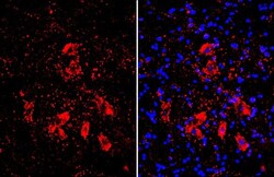
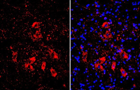
- ChAT Polyclonal Antibody detects Choline Acetyltransferase protein by immunohistochemical analysis. Sample: Frozen-sectioned mouse spinal cord. Red: Choline Acetyltransferase stained by ChAT Polyclonal Antibody (Product # PA5-29653) diluted at 1:300. Blue: Hoechst 33342 staining.
- Submitted by
- Invitrogen Antibodies (provider)
- Main image
- Experimental details
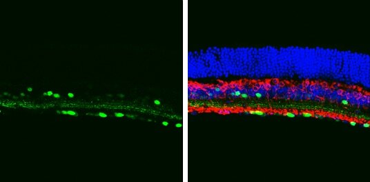
- ChAT Polyclonal Antibody detects Choline Acetyltransferase protein at cytoplasm and nucleus by immunohistochemical analysis. Sample: Paraffin-embedded mouse eye. Green: Choline Acetyltransferase stained by ChAT Polyclonal Antibody (Product # PA5-29653) diluted at 1:1,000. Red: beta Tubulin 3/ Tuj1, a neural marker, stained by beta Tubulin 3/ Tuj1 antibody [GT11710] diluted at 1:500. Blue: Fluoroshield with DAPI. Antigen Retrieval: Citrate buffer, pH 6.0, 15 min.

- Submitted by
- Invitrogen Antibodies (provider)
- Main image
- Experimental details
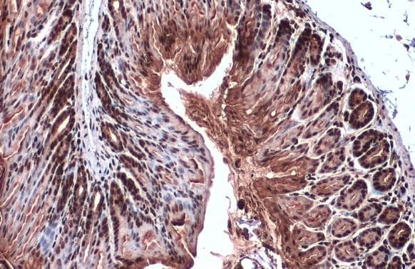
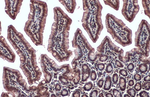
- Immunohistochemistry (Paraffin) analysis of ChAT was performed in paraffin-embedded mouse intestine tissue using ChAT Polyclonal Antibody (Product # PA5-29653) at a dilution of 1:500. Antigen Retrieval: Citrate buffer, pH 6.0, 15 min.
- Submitted by
- Invitrogen Antibodies (provider)
- Main image
- Experimental details
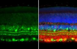
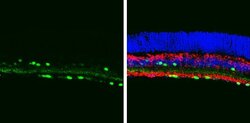
- Immunohistochemistry (Frozen) analysis of ChAT was performed in frozen sectioned adult mouse retina tissue using ChAT Polyclonal Antibody (Product # PA5-29653) at a dilution of 1:250 (Green). Red: Protein kinase C alpha staining. Blue: Fluoroshield with DAPI. Antigen Retrieval: Citrate buffer, pH 6.0, 15 min.
Supportive validation
- Submitted by
- Invitrogen Antibodies (provider)
- Main image
- Experimental details
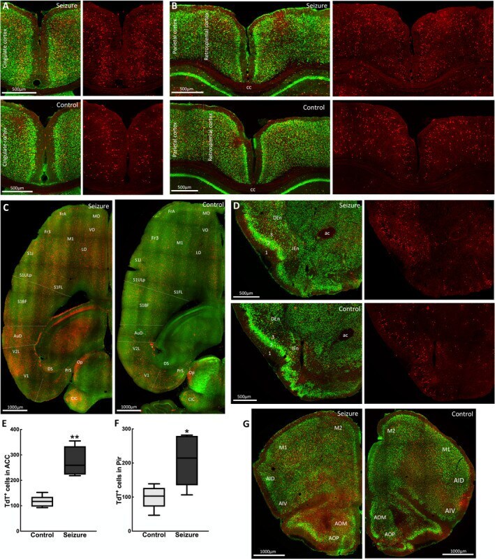
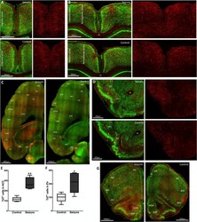
- FIGURE 3 Multiple cortical structures are activated during a focal impaired awareness seizure. (A) Coronal sections at bregma 0.13 mm showing TRAPed neurons in the anterior cingulate cortex during a seizure (top panel) and in controls (bottom panel) in merged images showing NeuN immunolabels in green and TdT + cells in red. (B) Coronal sections at bregma -1.91 mm showing TRAPed neurons in the retrosplenial and parietal cortices during a seizure (top panel) and in controls (bottom panel). (C) Horizontal sections at bregma -1.68 mm showing TdT activation in the frontal, orbital, motor, somatosensory, and visual cortices with (left panel) or without (right panel) a seizure. Merged images show TdT + cells (red) and ChAT immunolabeling (green). (D) Coronal sections at bregma 1.09 mm showing TRAPed neurons in the piriform (layers 1, 2, and 3) and endopiriform cortex. (E) A greater number of TdT-labeled cells were present in the ACC during a behavioral grade 2 seizure compared with controls (control 166 +- 10, seizure 274 +- 26; n = 5 mice per group, t( 5.22) = 5.65, P = .002). (F) More piriform cortex cell body layer II neurons were TdT-labeled compared with controls (control 100 +- 15, seizure 208 +- 33; n = 5 mice per group, t (5.56) = 2.91, P = .028). (G) Coronal sections at bregma 2.09 mm showing TRAPed neurons in motor and insular cortices, and olfactory nucleus. Regions marked are ac, anterior commissure; ACC, anterior cingulate cortex; AI, agranular insular cortex; AO, anter