Explore
 Validate
 Learn
Learn Immunohistochemistry
Antibody data
- Antibody Data
- Antigen structure
- References [3]
- Comments [0]
- Validations
- Immunohistochemistry [1]
Submit
Validation data
Reference
Comment
Report error
- Product number
- HPA015234 - Provider product page

- Provider
- Atlas Antibodies
- Proper citation
- Atlas Antibodies Cat#HPA015234, RRID:AB_1847604
- Product name
- Anti-PREX2
- Antibody type
- Polyclonal
- Description
- Polyclonal Antibody against Human PREX2, Gene description: phosphatidylinositol-3,4,5-trisphosphate-dependent Rac exchange factor 2, Alternative Gene Names: DEP.2, DEPDC2, FLJ12987, P-REX2, PPP1R129, Validated applications: IHC, Uniprot ID: Q70Z35, Storage: Store at +4°C for short term storage. Long time storage is recommended at -20°C.
- Reactivity
- Human
- Host
- Rabbit
- Conjugate
- Unconjugated
- Isotype
- IgG
- Vial size
- 100 µl
- Concentration
- 0.2 mg/ml
- Storage
- Store at +4°C for short term storage. Long time storage is recommended at -20°C.
- Handling
- The antibody solution should be gently mixed before use.
Submitted references Comparative Transcriptomic Analysis of Archival Human Vestibular Schwannoma Tissue from Patients with and without Tinnitus.
P-Rex2 mediation of synaptic plasticity contributes to bone cancer pain
Somatic mutations of PREX2 gene in patients with hepatocellular carcinoma
Bommakanti K, Seist R, Kukutla P, Cetinbas M, Batts S, Sadreyev RI, Stemmer-Rachamimov A, Brenner GJ, Stankovic KM
Journal of clinical medicine 2023 Apr 1;12(7)
Journal of clinical medicine 2023 Apr 1;12(7)
P-Rex2 mediation of synaptic plasticity contributes to bone cancer pain
Fu Q, Huang X, Wan S, Li Y, Li X, Su S, Xu X, Wu Y
Molecular Pain 2022;18
Molecular Pain 2022;18
Somatic mutations of PREX2 gene in patients with hepatocellular carcinoma
Yang M, Yen C, Chen Y, Fang C, Li C, Lee K, Lin Y, Weng C, Liu T, Huang S, Teh B, Chen Y
Scientific Reports 2019;9(1)
Scientific Reports 2019;9(1)
No comments: Submit comment
Supportive validation
- Submitted by
- Atlas Antibodies (provider)
- Enhanced method
- Orthogonal validation
- Main image
- Experimental details
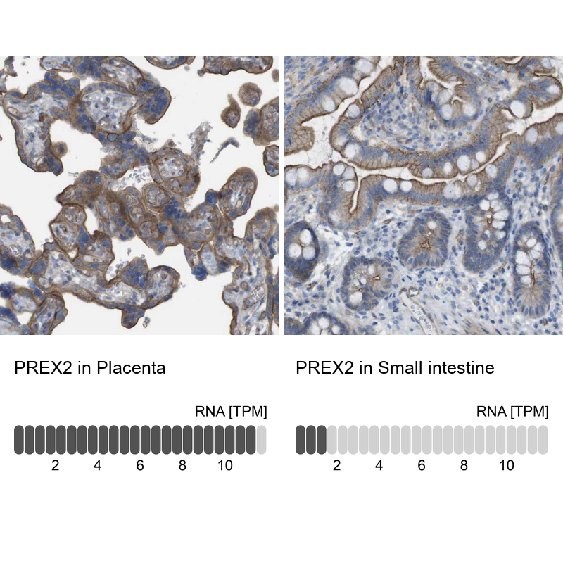
- Immunohistochemistry analysis in human placenta and small intestine tissues using HPA015234 antibody. Corresponding PREX2 RNA-seq data are presented for the same tissues.
- Sample type
- Human
- Protocol
- Protocol