 Explore
 Validate
 Learn
Learn Western blot
 Immunocytochemistry
Antibody data
- Antibody Data
- Antigen structure
- References [0]
- Comments [0]
- Validations
- Immunocytochemistry [7]
- Immunohistochemistry [3]
- Flow cytometry [2]
Submit
Validation data
Reference
Comment
Report error
- Product number
- MA5-34741 - Provider product page

- Provider
- Invitrogen Antibodies
- Product name
- MYST1 Recombinant Rabbit Monoclonal Antibody (JG36-05)
- Antibody type
- Monoclonal
- Antigen
- Recombinant full-length protein
- Description
- Positive Control: MCF-7, SH-SY-5Y, SiHa, human cervix tissue, human cervix cancer tissue, rat cervix tissue, K562.
- Reactivity
- Human, Mouse
- Host
- Rabbit
- Isotype
- IgG
- Antibody clone number
- JG36-05
- Vial size
- 100 μL
- Concentration
- 1 mg/mL
- Storage
- -20°C, Avoid Freeze/Thaw Cycles, store in dark
No comments: Submit comment
Supportive validation
- Submitted by
- Invitrogen Antibodies (provider)
- Main image
- Experimental details
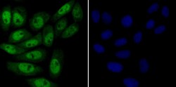
- Immunofluorescent analysis of KAT8 in SiHa cells (green). Samples were fixed in paraformaldehyde and permeabilised with 0.25% Triton X100/PBS, incubated with KAT8 monoclonal antibody (Product # MA5-34741), followed by DAPI (blue).
- Submitted by
- Invitrogen Antibodies (provider)
- Main image
- Experimental details
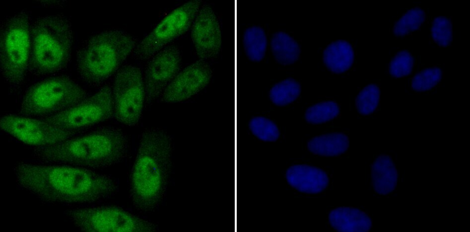
- Immunofluorescent analysis of KAT8 in SH-SY-5Y cells (green). Samples were fixed in paraformaldehyde and permeabilised with 0.25% Triton X100/PBS, incubated with KAT8 monoclonal antibody (Product # MA5-34741), followed by DAPI (blue).
- Submitted by
- Invitrogen Antibodies (provider)
- Main image

- Experimental details
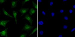
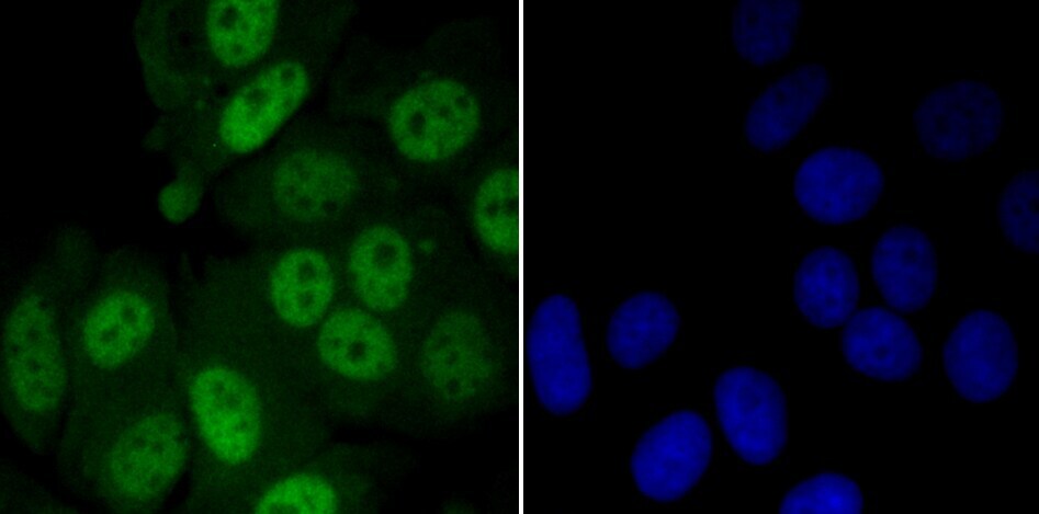
- Immunofluorescent analysis of KAT8 in MCF-7 cells (green). Samples were fixed in paraformaldehyde and permeabilised with 0.25% Triton X100/PBS, incubated with KAT8 monoclonal antibody (Product # MA5-34741), followed by DAPI (blue).

- Submitted by
- Invitrogen Antibodies (provider)
- Main image

- Experimental details
- Immunofluorescent analysis of KAT8 in MCF-7 cells (green). Samples were fixed in paraformaldehyde and permeabilised with 0.25% Triton X100/PBS, incubated with KAT8 monoclonal antibody (Product # MA5-34741), followed by DAPI (blue).
- Submitted by
- Invitrogen Antibodies (provider)
- Main image

- Experimental details
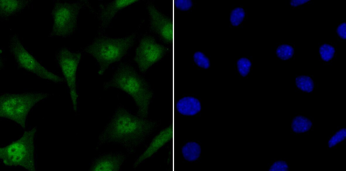
- Immunofluorescent analysis of KAT8 in SH-SY-5Y cells (green). Samples were fixed in paraformaldehyde and permeabilised with 0.25% Triton X100/PBS, incubated with KAT8 monoclonal antibody (Product # MA5-34741), followed by DAPI (blue).

- Submitted by
- Invitrogen Antibodies (provider)
- Main image

- Experimental details
- Immunofluorescent analysis of KAT8 in SiHa cells (green). Samples were fixed in paraformaldehyde and permeabilised with 0.25% Triton X100/PBS, incubated with KAT8 monoclonal antibody (Product # MA5-34741), followed by DAPI (blue).

- Submitted by
- Invitrogen Antibodies (provider)
- Main image
- Experimental details
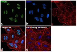
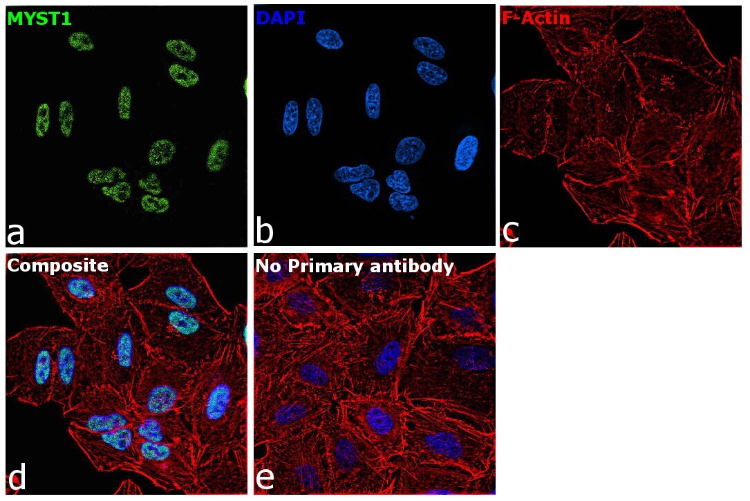
- Immunofluorescence analysis of MYST1 was performed using 70% confluent log phase A549 cells. The cells were fixed with 4% paraformaldehyde for 15 minutes, permeabilized with 0.1% Triton™ X-100 for 15 minutes, and blocked with 2% BSA for 45 minutes at room temperature. The cells were labeled with MYST1 Recombinant Rabbit Monoclonal Antibody (JG36-05) (Product # MA5-34741) at 1:200 dilution in 0.1% BSA, incubated at 4 degree celsius overnight and then labeled with Donkey anti-Rabbit IgG (H+L) Highly Cross-Adsorbed Secondary Antibody, Alexa Fluor™ Plus 488 (Product # A32790), (1:3,000), for 45 minutes at room temperature (Panel a: Green). Nuclei (Panel b: Blue) were stained with ProLong™ Diamond Antifade Mountant with DAPI (Product # P36962). F-actin (Panel c: Red) was stained with Rhodamine Phalloidin (Product # R415, 1:300). Panel d represents the merged image showing nuclear localization. Panel e represents control cells with no primary antibody to assess background. The images were captured at 60X magnification.
Supportive validation
- Submitted by
- Invitrogen Antibodies (provider)
- Main image

- Experimental details
- Immunohistochemistry analysis of KAT8 in paraffin-embedded rat cervix tissue. Samples were incubated with KAT8 monoclonal antibody (Product # MA5-34741), and followed by hematoxylin.
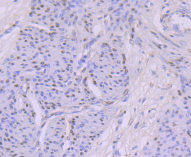
- Submitted by
- Invitrogen Antibodies (provider)
- Main image
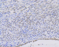
- Experimental details
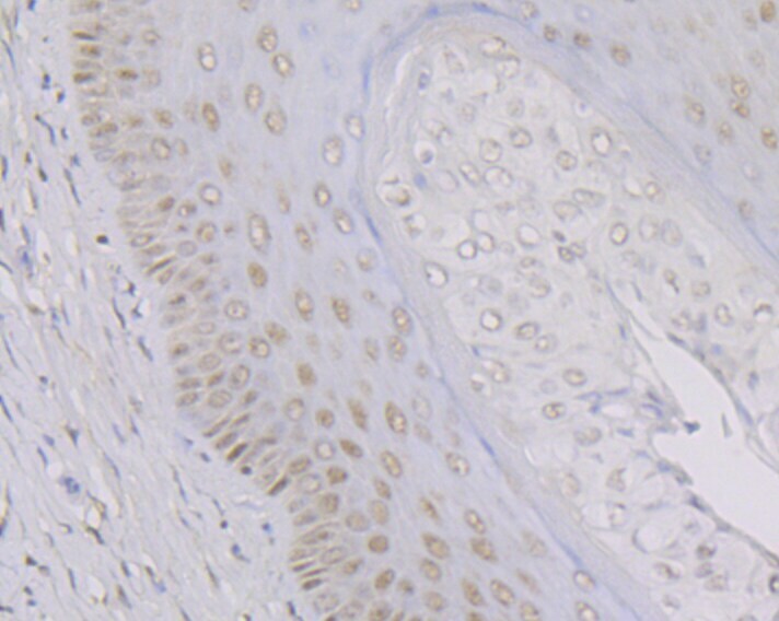
- Immunohistochemistry analysis of KAT8 in paraffin-embedded human cervix cancer tissue. Samples were incubated with KAT8 monoclonal antibody (Product # MA5-34741), and followed by hematoxylin.
- Submitted by
- Invitrogen Antibodies (provider)
- Main image
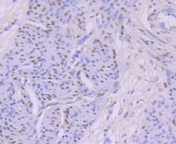
- Experimental details
- Immunohistochemistry analysis of KAT8 in paraffin-embedded human cervix tissue. Samples were incubated with KAT8 monoclonal antibody (Product # MA5-34741), and followed by hematoxylin.
Supportive validation
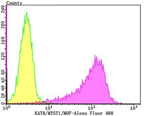
- Submitted by
- Invitrogen Antibodies (provider)
- Main image

- Experimental details
- Flow cytometry of KAT8 in K562 cells (yellow) compared with an unlabelled control (cells without incubation with primary antibody; purple). Samples were incubated with KAT8 monoclonal antibody (Product # MA5-34741) at a dilution of 1:100, followed by Alexa Fluor 488-conjugated goat anti-rabbit IgG.
- Submitted by
- Invitrogen Antibodies (provider)
- Main image
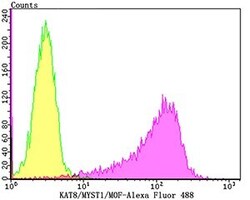
- Experimental details
- Flow cytometry of KAT8 in K562 cells (yellow) compared with an unlabelled control (cells without incubation with primary antibody; purple). Samples were incubated with KAT8 monoclonal antibody (Product # MA5-34741) at a dilution of 1:100, followed by Alexa Fluor 488-conjugated goat anti-rabbit IgG.
