Explore
 Validate
 Learn
Learn Western blot
 Immunocytochemistry
 Immunohistochemistry
Antibody data
- Antibody Data
- Antigen structure
- References [1]
- Comments [0]
- Validations
- Immunocytochemistry [2]
- Other assay [1]
Submit
Validation data
Reference
Comment
Report error
- Product number
- MA5-27662 - Provider product page

- Provider
- Invitrogen Antibodies
- Product name
- VAChT Monoclonal Antibody (S6)
- Antibody type
- Monoclonal
- Antigen
- Synthetic peptide
- Description
- A dilution of 1:50-1:200 of MA5-27662 was sufficient for detection of VAChT Transporter in rat brain using immunohistochemistry analysis and goat anti-mouse IgG:HRP as the secondary antibody. Detects approximately 56kDa. This antibody was formerly sold as S6-38.
- Reactivity
- Human, Mouse, Rat
- Host
- Mouse
- Isotype
- IgG
- Antibody clone number
- S6
- Vial size
- 100 μg
- Concentration
- 1 mg/mL
- Storage
- -20°C
Submitted references Lack of Mucosal Cholinergic Innervation Is Associated With Increased Risk of Enterocolitis in Hirschsprung's Disease.
Keck S, Galati-Fournier V, Kym U, Moesch M, Usemann J, Müller I, Subotic U, Tharakan SJ, Krebs T, Stathopoulos E, Schmittenbecher P, Cholewa D, Romero P, Reingruber B, Bruder E, Group NS, Holland-Cunz S
Cellular and molecular gastroenterology and hepatology 2021;12(2):507-545
Cellular and molecular gastroenterology and hepatology 2021;12(2):507-545
No comments: Submit comment
Supportive validation
- Submitted by
- Invitrogen Antibodies (provider)
- Main image

- Experimental details
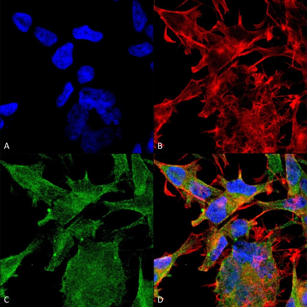
- Immunofluorescent analysis of VAChT in human neuroblastoma cell line (SK-N-BE). Sample was fixed with 4% formaldehyde (15 min at RT), incubated with VAChT monoclonal antibody (Product # MA5-27662) using a dilution of 1:100 (1 hour at RT), and followed by Goat Anti-Mouse 488, Phalloidin Texas Red and DAPI secondary antibody at a dilution of 1:200, 1:1000 (60 min at RT) and 1:5000 (5 min at RT). Images are shown as follows: (A) DAPI (blue) nuclear stain, B) Phalloidin Texas Red F-Actin stain, C) VAChT Antibody, D) Merged image. Magnification: 60x.
- Submitted by
- Invitrogen Antibodies (provider)
- Main image
- Experimental details
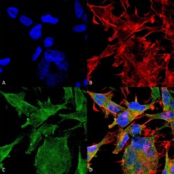
- Immunofluorescent analysis of VAChT in human neuroblastoma cell line (SK-N-BE). Sample was fixed with 4% formaldehyde (15 min at RT), incubated with VAChT monoclonal antibody (Product # MA5-27662) using a dilution of 1:100 (1 hour at RT), and followed by Goat Anti-Mouse 488, Phalloidin Texas Red and DAPI secondary antibody at a dilution of 1:200, 1:1000 (60 min at RT) and 1:5000 (5 min at RT). Images are shown as follows: (A) DAPI (blue) nuclear stain, B) Phalloidin Texas Red F-Actin stain, C) VAChT Antibody, D) Merged image. Magnification: 60x.

Supportive validation
- Submitted by
- Invitrogen Antibodies (provider)
- Main image
- Experimental details
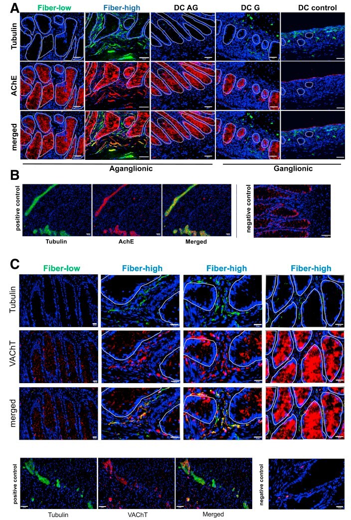
- Figure 4 Cholinergic innervation in distal colon of HSCR patients. ( A ) Immunofluorescence of epithelial/mucosal region from aganglionic and ganglionic colonic tissue from HSCR and control patients using tubulin (Alexa647, green ) and AChE (Cy3, red ). DAPI ( blue ) shows cell nuclei. Encircled areas mark the epithelial crypt/lumen. Scale bar: 50 mum. DC control images: scale bar: 100 mum. DC AG: aganglionic descending colon; DC G: ganglionic descending colon; DC control: ganglionic descending colon from control patients. ( B ) Positive control: immunofluorescence of myenteric plexus ganglia from ganglionic control tissue. Negative control: immunofluorescence of epithelial/mucosal region from aganglionic fiber-high colonic tissue using secondary antibody controls mouse IgG2b-biotin/SA-Cy3 together with mouse IgG2a-A647. Scale bar: 50 mum. ( C ) Immunofluorescence of epithelial/mucosal region from aganglionic fiber-low and fiber-high colonic tissue from HSCR patients using tubulin (Alexa647, green ) and VAChT (A555, red ). DAPI ( blue ) shows cell nuclei. Encircled areas mark the epithelial crypt/lumen. Positive control: immunofluorescence of myenteric plexus ganglia from ganglionic control tissue. Scale bar: 50 mum. Negative control: immunofluorescence of epithelial/mucosal region from aganglionic fiber-high colonic tissue using secondary antibody controls mouse IgG1-A555 together with mouse IgG2a-A647. Scale bar: 20 mum.