Explore
 Validate
 Learn
Learn Western blot
 ELISA
ELISAAntibody data
- Antibody Data
- Antigen structure
- References [0]
- Comments [0]
- Validations
- Western blot [1]
- Immunocytochemistry [1]
- Immunohistochemistry [1]
Submit
Validation data
Reference
Comment
Report error
- Product number
- 600-401-MK7 - Provider product page

- Provider
- Invitrogen Antibodies
- Product name
- VAChT Polyclonal Antibody
- Antibody type
- Polyclonal
- Antigen
- Synthetic peptide
- Reactivity
- Human, Rat
- Host
- Rabbit
- Isotype
- IgG
- Vial size
- 100 µg
- Concentration
- 0.9 mg/mL
- Storage
- -20° C, Avoid Freeze/Thaw Cycles
No comments: Submit comment
Supportive validation
- Submitted by
- Invitrogen Antibodies (provider)
- Main image
- Experimental details
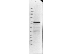
- Western Blot of Rabbit Anti-VAChT Antibody. Molecular Weight Marker: Opal Pre-stained (p/n MB-210-0500). Lane 1: Rat brain, post natal, Whole Cell Lysate (p/n W12-001-MQ6). Primary Antibody: Anti-VAChT Antibody at 1:1000 overnight at 4°C. Secondary Antibody: Donkey Anti-Rabbit IgG HRP (p/n 611-703-127) at 1:70,000 for 1hr at RT. Blocking: BlockOut Universal Buffer (p/n MB-073). Expected: ~57 kDa, protein glycosylation.

Supportive validation
- Submitted by
- Invitrogen Antibodies (provider)
- Main image
- Experimental details
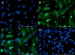
- Immunofluorescence of Rabbit Anti-VAChT Antibody. Cell Line: Post-natal rat pup (PND1) heterogeneous brain cells. Primary Antibody: Rabbit Anti-VAChT Antibody at 15µg/mL overnight at 4°C. Counterstain Antibody: Donkey Anti-Rabbit IgG DyLight™ 488 Conjugated (p/n 611-741-127) at 5µg/mL for 1hr at RT. Fixative: 4% PFA. Permeabilization: 0.3% Triton X-100. Nuclear Counter Stain: DAPI. Image: A) DAPI (blue). B) Anti-VAChT (green) and Secondary. C) Merge of A+B images. D) Secondary only. Localization: predicted membrane, Golgi apparatus. Antibody detecting dendrites and axons and specific organelles in the nucleus (nucleolus). Staining correspondent to confirmed Slc18a3 staining in mouse brain P7 Gensat project: http://www.gensat.org/imagenavigator.jsp?imageID=66611
Supportive validation
- Submitted by
- Invitrogen Antibodies (provider)
- Main image
- Experimental details
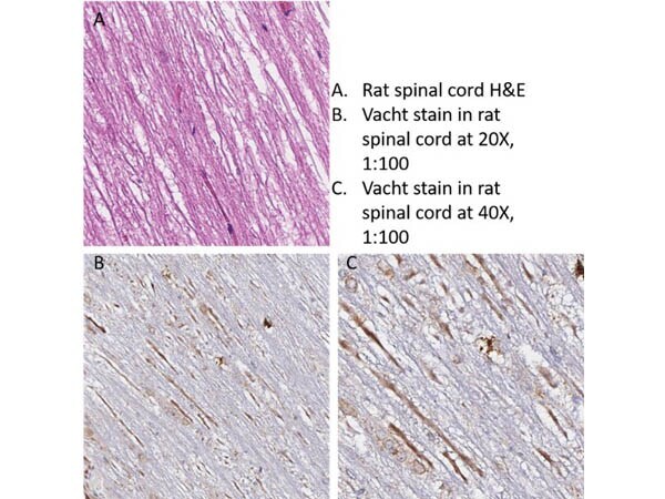
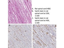
- Immunohistochemistry of Rabbit Anti-VAChT Antibody. Tissue: Rat Spinal Cord. Primary Antibody: Rabbit Anti-VAChT Antibody at 1:100 for 30min at RT. Secondary Antibody: Anti-Rabbit Poly-HRP-IgG. Ready to use 8 min at RT. Antigen Retrieval: HIER using Citrate Buffer for 20 min. Counterstain: Hematoxylin.