Explore
 Validate
 Learn
Learn Immunocytochemistry
Antibody data
- Antibody Data
- Antigen structure
- References [3]
- Comments [0]
- Validations
- Immunocytochemistry [1]
- Flow cytometry [1]
- Other assay [2]
Submit
Validation data
Reference
Comment
Report error
- Product number
- MA5-18149 - Provider product page

- Provider
- Invitrogen Antibodies
- Product name
- CD63 Monoclonal Antibody (MEM-259), Alexa Fluor™ 488
- Antibody type
- Monoclonal
- Antigen
- Other
- Description
- This antibody reacts with an extracellular/luminal epitope of CD63 (LAMP-3), a 40-60 kDa tetraspan glycoprotein expressed by granulocytes, platelets, T cells, monocytes/macrophages and endothelial cells. Cell surface exposition of CD63 is usually activation-dependent.
- Reactivity
- Human
- Host
- Mouse
- Conjugate
- Green dye
- Isotype
- IgG
- Antibody clone number
- MEM-259
- Vial size
- 100 Tests
- Storage
- 4° C, store in dark, DO NOT FREEZE!
Submitted references Irreversible alteration of extracellular vesicle and cell-free messenger RNA profiles in human plasma associated with blood processing and storage.
Nondestructive production of exosomes loaded with ultrathin palladium nanosheets for targeted bio-orthogonal catalysis.
Microenvironmental pH and Exosome Levels Interplay in Human Cancer Cell Lines of Different Histotypes.
Kim HJ, Rames MJ, Tassi Yunga S, Armstrong R, Morita M, Ngo ATP, McCarty OJT, Civitci F, Morgan TK, Ngo TTM
Scientific reports 2022 Feb 8;12(1):2099
Scientific reports 2022 Feb 8;12(1):2099
Nondestructive production of exosomes loaded with ultrathin palladium nanosheets for targeted bio-orthogonal catalysis.
Sebastian V, Sancho-Albero M, Arruebo M, Pérez-López AM, Rubio-Ruiz B, Martin-Duque P, Unciti-Broceta A, Santamaría J
Nature protocols 2021 Jan;16(1):131-163
Nature protocols 2021 Jan;16(1):131-163
Microenvironmental pH and Exosome Levels Interplay in Human Cancer Cell Lines of Different Histotypes.
Logozzi M, Mizzoni D, Angelini DF, Di Raimo R, Falchi M, Battistini L, Fais S
Cancers 2018 Oct 5;10(10)
Cancers 2018 Oct 5;10(10)
No comments: Submit comment
Supportive validation
- Submitted by
- Invitrogen Antibodies (provider)
- Main image

- Experimental details
- Immunofluorescent analysis of CD63 in human primary fibroblasts (green) using a CD63 monoclonal antibody (Product # MA5-18149). Cytoplasmic actin was stained with a fluorescent red phalloidin and nuclei were stained with DAPI (blue).
- Conjugate
- Green dye

Supportive validation
- Submitted by
- Invitrogen Antibodies (provider)
- Main image

- Experimental details
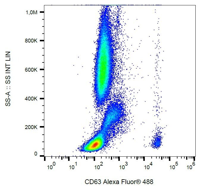
- Flow cytometry analysis of IgE-activated peripheral human blood stained with anti-human CD63 (MEM-259) Alexa Fluor® 488 Monoclonal antibody (Product # MA5-18149).
- Conjugate
- Green dye
Supportive validation
- Submitted by
- Invitrogen Antibodies (provider)
- Main image
- Experimental details
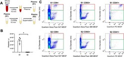
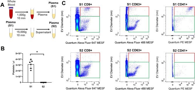
- Figure 3 Effect of differential centrifugation on EVs using flow cytometry. ( A ) Schematic diagram of differentially processed plasma using single spin (S1: 1000 x g centrifugation) and double spin (S2: 15,000 x g secondary spin after the initial single spin S1). ( B ) Platelet concentration in differentially processed plasma from three healthy individuals (n = 3) was measured in independent technical replicates using a haemocytometer. The error bar represented standard deviations for the indicated blood processing conditions. P -value was calculated using Wilcoxon test (* P < 0.05). ( C ) Representative flow cytometry dot plot of EV diameter (nm) versus fluorescent intensity in Quantum Alexa Fluor MESF units for S1 and S2 using FlowJo. Quantum Alexa Fluor 647 MESF was used for Alexa Fluor 647 conjugated CD9 stained plasma, Quantum Alexa Fluor 488 MESF was used for Alexa Fluor 488 conjugated CD63 stained plasma, and Quantum PE MESF was used for PE conjugated CD41 stained plasma. Events were gated into two subpopulations: 150 to 1000 nm (green box) and from 1000 to 3000 nm (red box).
- Conjugate
- Green dye
- Submitted by
- Invitrogen Antibodies (provider)
- Main image
- Experimental details
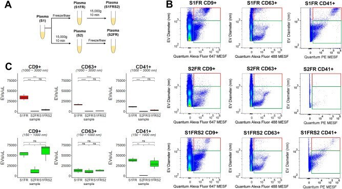
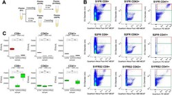
- Figure 5 Effect of post-thaw processing on EVs using flow cytometry. ( A ) Schematic diagram of differentially processed plasma (S1, S2), respective freeze thaw samples (S1FR, S2FR), and secondary spin after post-freeze/thaw plasma S1FR (S1FRS2). ( B ) Representative flow cytometry dot plot of EV diameter (nm) versus fluorescent intensity in Quantum MESF units for CD9 + EVs, CD63 + , and CD41 + EVs in S1FR, S2FR, and S1FRS2 conditions using FlowJo. Quantum Alexa Fluor 647 MESF is used for Alexa Fluor 647 conjugated CD9 stained plasma, Quantum Alexa Fluor 488 MESF is used for Alexa Fluor 488 conjugated CD63 stained plasma, and Quantum PE MESF is used for PE conjugated CD41 stained plasma. Events were gated from 150 to 1000 nm (green box) and from 1000 to 3000 nm (red box). ( C ) Box plot of CD9 + , CD63 + , CD41 + EV concentration from 1000-3000 nm (red) and 150-1000 nm (green) for S1FR, S2FR, and S1FRS2 using R. Statistical significance were obtained from three healthy volunteers for each freeze thaw processing condition using Tukey's multiple comparisons (ns = not significant, P > 0.05; * P < 0.05, *** P < 0.001, **** P < 0.0001).
- Conjugate
- Green dye