Explore
 Validate
 Learn
Learn Immunohistochemistry
Antibody data
- Antibody Data
- Antigen structure
- References [1]
- Comments [0]
- Validations
- Immunohistochemistry [1]
- Flow cytometry [1]
Submit
Validation data
Reference
Comment
Report error
- Product number
- MAB5417 - Provider product page

- Provider
- R&D Systems
- Product name
- Mouse CD63 Antibody
- Antibody type
- Monoclonal
- Description
- Protein A or G purified from hybridoma culture supernatant. Detects mouse CD63 in direct ELISAs.
- Reactivity
- Mouse
- Host
- Rat
- Conjugate
- Unconjugated
- Antigen sequence
P41731- Isotype
- IgG
- Antibody clone number
- 446703
- Vial size
- 100 ug
- Concentration
- LYOPH
- Storage
- Use a manual defrost freezer and avoid repeated freeze-thaw cycles. 12 months from date of receipt, -20 to -70 °C as supplied. 1 month, 2 to 8 °C under sterile conditions after reconstitution. 6 months, -20 to -70 °C under sterile conditions after reconstitution.
Submitted references Bacterial colonization of host cells in the absence of cholesterol.
Gilk SD, Cockrell DC, Luterbach C, Hansen B, Knodler LA, Ibarra JA, Steele-Mortimer O, Heinzen RA
PLoS pathogens 2013 Jan;9(1):e1003107
PLoS pathogens 2013 Jan;9(1):e1003107
No comments: Submit comment
Supportive validation
- Submitted by
- R&D Systems (provider)
- Main image

- Experimental details
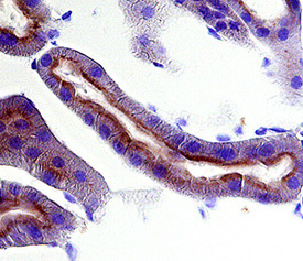
- CD63 in Mouse Kidney. CD63 was detected in perfusion fixed frozen sections of mouse kidney using Rat Anti-Mouse CD63 Monoclonal Antibody (Catalog # MAB5417) at 25 µg/mL overnight at 4 °C. Tissue was stained using the Anti-Rat HRP-DAB Cell & Tissue Staining Kit (brown; Catalog # CTS017) and counterstained with hematoxylin (blue). Specific labeling was localized to the plasma membrane of epithelial cells in convoluted tubules. View our protocol for Chromogenic IHC Staining of Frozen Tissue Sections.
Supportive validation
- Submitted by
- R&D Systems (provider)
- Main image
- Experimental details
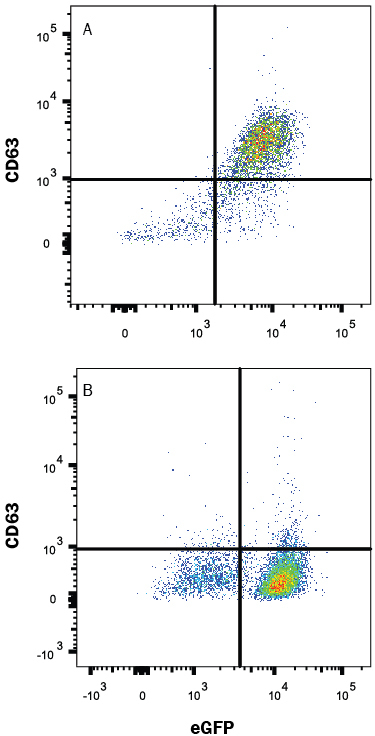
- Detection of CD63 in HEK293 Human Cell Line Transfected with Mouse CD63 and eGFP by Flow Cytometry. HEK293 human embryonic kidney cell line transfected with either (A) mouse CD63 or (B) irrelevant transfectants and eGFP were stained with Rat Anti-Mouse CD63 Monoclonal Antibody (Catalog # MAB5417) followed by Allophycocyanin-conjugated Anti-Rat IgG Secondary Antibody (Catalog # F0113). Quadrant markers were set based on control antibody staining (Catalog # MAB005). View our protocol for Staining Membrane-associated Proteins.