Explore
 Validate
 Learn
Learn Western blot
 ELISA
ELISA Immunocytochemistry
Antibody data
- Antibody Data
- Antigen structure
- References [3]
- Comments [0]
- Validations
- Immunocytochemistry [4]
- Immunohistochemistry [3]
- Other assay [1]
Submit
Validation data
Reference
Comment
Report error
- Product number
- PA5-92370 - Provider product page

- Provider
- Invitrogen Antibodies
- Product name
- CD63 Polyclonal Antibody
- Antibody type
- Polyclonal
- Antigen
- Recombinant full-length protein
- Description
- Immunogen sequence: AGYVFRDKVM SEFNNNFRQQ MENYPKNNHT ASILDRMQAD FKCCGAANYT DWEKIPSMSK NRVPDSCCIN VTVGCGINFN EKAIHKEGCV EKIGGWLRKN V; Positive Samples: A375, 22Rv1, Mouse kidney, Mouse spleen; Cellular Location: Cell membrane, Endosome, Late endosome membrane, Lysosome membrane, Melanosome, Multi-pass membrane protein, multivesicular body
- Reactivity
- Human
- Host
- Rabbit
- Isotype
- IgG
- Vial size
- 100 μL
- Concentration
- 1.07 mg/mL
- Storage
- -20°C, Avoid Freeze/Thaw Cycles
Submitted references Sustained release of exosomes loaded into polydopamine-modified chitin conduits promotes peripheral nerve regeneration in rats.
A Novel Localization in Human Large Extracellular Vesicles for the EGF-CFC Founder Member CRIPTO and Its Biological and Therapeutic Implications.
Macrophage secretion of miR-106b-5p causes renin-dependent hypertension.
Li C, Liu SY, Zhang M, Pi W, Wang B, Li QC, Lu CF, Zhang PX
Neural regeneration research 2022 Sep;17(9):2050-2057
Neural regeneration research 2022 Sep;17(9):2050-2057
A Novel Localization in Human Large Extracellular Vesicles for the EGF-CFC Founder Member CRIPTO and Its Biological and Therapeutic Implications.
Mantile F, Kisovec M, Adamo G, Romancino DP, Hočevar M, Božič D, Bedina Zavec A, Podobnik M, Stoppelli MP, Kisslinger A, Bongiovanni A, Kralj-Iglič V, Liguori GL
Cancers 2022 Jul 29;14(15)
Cancers 2022 Jul 29;14(15)
Macrophage secretion of miR-106b-5p causes renin-dependent hypertension.
Oh J, Matkovich SJ, Riek AE, Bindom SM, Shao JS, Head RD, Barve RA, Sands MS, Carmeliet G, Osei-Owusu P, Knutsen RH, Zhang H, Blumer KJ, Nichols CG, Mecham RP, Baldán Á, Benitez BA, Sequeira-Lopez ML, Gomez RA, Bernal-Mizrachi C
Nature communications 2020 Sep 23;11(1):4798
Nature communications 2020 Sep 23;11(1):4798
No comments: Submit comment
Supportive validation
- Submitted by
- Invitrogen Antibodies (provider)
- Main image
- Experimental details
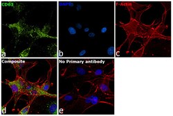
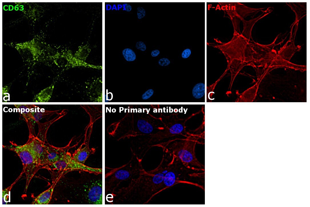
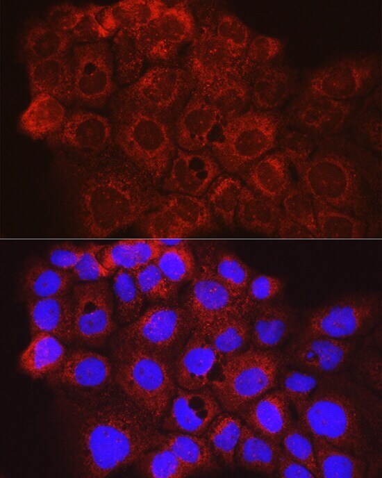
- Immunofluorescence analysis of CD63 was performed using 70% confluent log phase U-87 MG cells. The cells were fixed with 4% paraformaldehyde for 10 minutes, permeabilized with 0.1% Triton™ X-100 for 15 minutes, and blocked with 2% BSA for 45 minutes at room temperature. The cells were labeled with CD63 Polyclonal Antibody (Product # PA5-92370) at 1:100 dilution in 0.1% BSA, incubated at 4 degrees celsius overnight, and then labeled with Donkey anti-Rabbit IgG (H+L) Highly Cross-Adsorbed Secondary Antibody, Alexa Fluor Plus 488 (Product # A32790, 1:2000 dilution), for 45 minutes at room temperature (Panel a: Green). Nuclei (Panel b: Blue) were stained with ProLong™ Diamond Antifade Mountant with DAPI (Product # P36962). F-actin (Panel c: Red) was stained with Rhodamine Phalloidin (Product # R415, 1:300). Panel d represents the merged image showing cytoplasmic (lysosomal pattern) localization. Panel e represents control cells with no primary antibody to assess the background. The images were captured at 60X magnification.
- Submitted by
- Invitrogen Antibodies (provider)
- Main image

- Experimental details
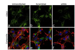
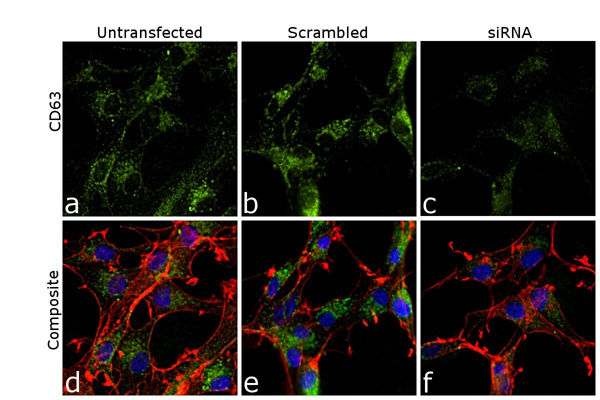
- Knockdown of CD63 was achieved by transfecting U-87 MG cells with CD63-specific siRNA (Silencer® select Product # S2700, S2699). Immunofluorescence analysis was performed on untransfected U-87 MG cells (panel a, d), transfected with non-specific scrambled siRNA (panels b, e), and transfected with CD63 specific siRNA (panels c,f). Cells were fixed, permeabilized, and labelled with CD63 Polyclonal Antibody (Product # PA5-92370, 1:100 dilution) followed by Donkey anti-Rabbit IgG (H+L) Highly Cross-Adsorbed Secondary Antibody, Alexa Fluor Plus 488 (Product # A32790, 1:2000 dilution). Nuclei (blue) were stained using ProLong™ Diamond Antifade Mountant with DAPI (Product # P36962), and Rhodamine Phalloidin (Product # R415, 1:300) was used for cytoskeletal F-actin (Red) staining. Reduction of specific signal was observed upon siRNA-mediated knockdown (panel c,f) confirming the specificity of the antibody to CD63 (Green). The Images were captured at 60X magnification.

- Submitted by
- Invitrogen Antibodies (provider)
- Main image
- Experimental details
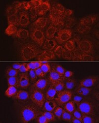
- Immunofluorescence analysis of CD63 in A431 cells. Samples were incubated with CD63 Polyclonal antibody (Product # PA5-92370) using a dilution of 1:100 (40x lens). Blue: DAPI for nuclear staining.
- Submitted by
- Invitrogen Antibodies (provider)
- Main image
- Experimental details
- Knockdown of CD63 was achieved by transfecting U-87 MG cells with CD63-specific siRNA (Silencer® select Product # S2700, S2699). Immunofluorescence analysis was performed on untransfected U-87 MG cells (panel a, d), transfected with non-specific scrambled siRNA (panels b, e), and transfected with CD63 specific siRNA (panels c,f). Cells were fixed, permeabilized, and labelled with CD63 Polyclonal Antibody (Product # PA5-92370, 1:100 dilution) followed by Donkey anti-Rabbit IgG (H+L) Highly Cross-Adsorbed Secondary Antibody, Alexa Fluor Plus 488 (Product # A32790, 1:2000 dilution). Nuclei (blue) were stained using ProLong™ Diamond Antifade Mountant with DAPI (Product # P36962), and Rhodamine Phalloidin (Product # R415, 1:300) was used for cytoskeletal F-actin (Red) staining. Reduction of specific signal was observed upon siRNA-mediated knockdown (panel c,f) confirming the specificity of the antibody to CD63 (Green). The Images were captured at 60X magnification.
Supportive validation
- Submitted by
- Invitrogen Antibodies (provider)
- Main image
- Experimental details
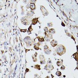
- Immunohistochemistry analysis of CD63 in paraffin-embedded human breast cancer using. Samples were incubated with CD63 Polyclonal antibody (Product # PA5-92370) using a dilution of 1:100 (40x lens). Perform high pressure antigen retrieval with 10 mM citrate buffer pH 6.0 before commencing with IHC staining protocol.

- Submitted by
- Invitrogen Antibodies (provider)
- Main image

- Experimental details
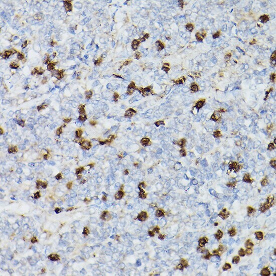
- Immunohistochemistry analysis of CD63 in paraffin-embedded human lymphoma. Samples were incubated with CD63 Polyclonal antibody (Product # PA5-92370) using a dilution of 1:100 (40x lens). Perform high pressure antigen retrieval with 10 mM citrate buffer pH 6.0 before commencing with IHC staining protocol.
- Submitted by
- Invitrogen Antibodies (provider)
- Main image

- Experimental details
- Immunohistochemistry analysis of CD63 in paraffin-embedded human lymphoma. Samples were incubated with CD63 Polyclonal antibody (Product # PA5-92370) using a dilution of 1:100 (40x lens). Perform high pressure antigen retrieval with 10 mM citrate buffer pH 6.0 before commencing with IHC staining protocol.

Supportive validation
- Submitted by
- Invitrogen Antibodies (provider)
- Main image
- Experimental details
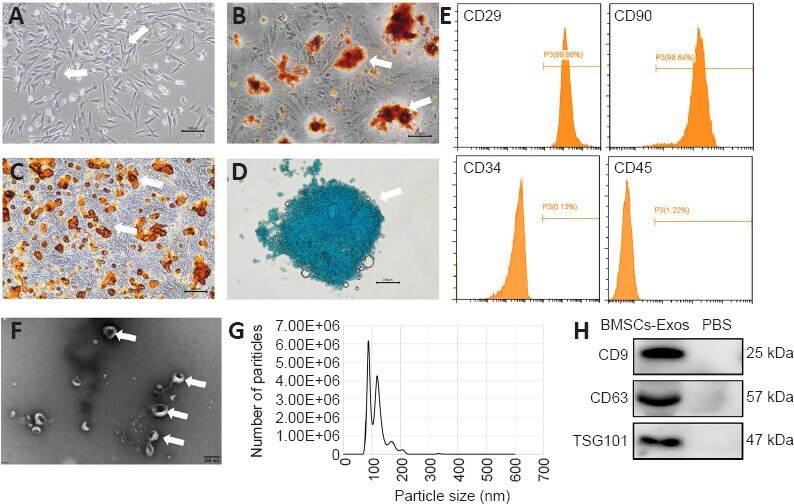
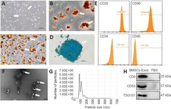
- Identification of BMSCs and BMSC-Exos . (A) Representative spindle shape (white arrows) of BMSCs. (B) Alizarin red staining shows calcium nodules (white arrows) after osteogenic differentiation. (C) Oil red O staining shows lipid droplets (white arrows) after adipogenic differentiation. (D) Alcian blue staining shows a chondrocyte group (white arrow) after chondrogenic differentiation. Scale bars: 100 mum (A-D). (E) Flow cytometric analysis of surface markers of BMSCs. The BMSCs were positive for CD29 and CD90 and negative for CD34 and CD45. (F) Representative morphology of BMSC-Exos (white arrows) observed by transmission electron microscopy. Scale bar: 200 nm. (G) Particle size distribution in purified BMSC-Exos measured by nanoparticle tracking analysis. (H) Western blot analysis of the specific exosome surface markers CD9, CD63, and TSG101. BMSCs: Bone mesenchymal stem cells; BMSCs-Exos: exosomes derived from bone mesenchymal stem cells; PBS: phosphate buffered saline; TSG101: tumor susceptibility gene 101.