Explore
 Validate
 Learn
Learn Western blot
 ELISA
ELISA Immunocytochemistry
Antibody data
- Antibody Data
- Antigen structure
- References [0]
- Comments [0]
- Validations
- Immunocytochemistry [5]
- Immunoprecipitation [1]
- Immunohistochemistry [2]
- Other assay [1]
Submit
Validation data
Reference
Comment
Report error
- Product number
- PA5-117019 - Provider product page

- Provider
- Invitrogen Antibodies
- Product name
- UBE2C Polyclonal Antibody
- Antibody type
- Polyclonal
- Antigen
- Recombinant full-length protein
- Description
- Positive Samples: 22Rv1, THP-1, U-87MG, SW620, SKOV3 Immunogen sequence: MASQNRDPAA TSVAAARKGA EPSGGAARGP VGKRLQQELM TLMMSGDKGI SAFPESDNLF KWVGTIHGAA GTVYEDLRYK LSLEFPSGYP YNAPTVKFLT PCYHPNVDTQ GNICLDILKE KWSALYDVRT ILLSIQSLLG EPNIDSPLNT HAAELWKNPT AFKKYLQETY SKQVTSQEP
- Reactivity
- Human, Mouse, Rat
- Host
- Rabbit
- Isotype
- IgG
- Vial size
- 100 μL
- Concentration
- 1.79 mg/mL
- Storage
- -20°C, Avoid Freeze/Thaw Cycles
No comments: Submit comment
Supportive validation
- Submitted by
- Invitrogen Antibodies (provider)
- Main image
- Experimental details
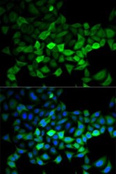
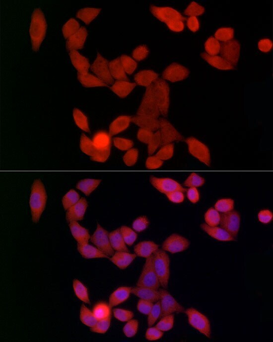
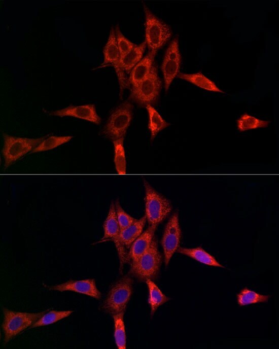
- Immunocytochemistry/Immunofluorescence analysis of UBE2C in HeLa cells using UBE2C Polyclonal Antibody (Product # PA5-117019) at a dilution of 1:100. Blue: DAPI for nuclear staining.
- Submitted by
- Invitrogen Antibodies (provider)
- Main image
- Experimental details
- Immunofluorescence analysis of UBE2C in HeLa cells. Samples were incubated with UBE2C Polyclonal antibody (Product # PA5-117019) using a dilution of 1:100 (40x lens). Blue: DAPI for nuclear staining.
- Submitted by
- Invitrogen Antibodies (provider)
- Main image
- Experimental details
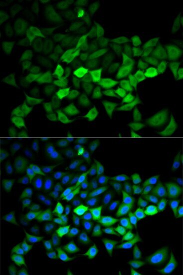
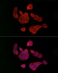
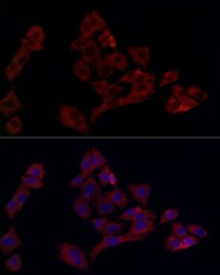
- Immunofluorescence analysis of UBE2C in MCF7 cells. Samples were incubated with UBE2C Polyclonal antibody (Product # PA5-117019) using a dilution of 1:100 (40x lens). Blue: DAPI for nuclear staining.
- Submitted by
- Invitrogen Antibodies (provider)
- Main image
- Experimental details
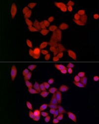
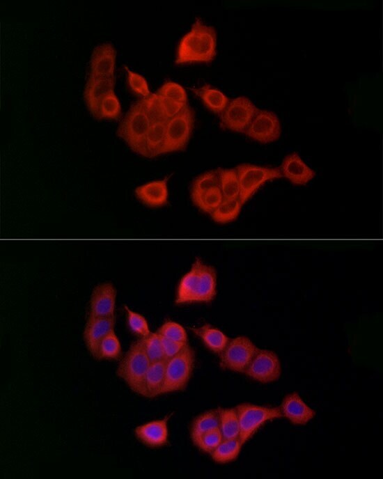
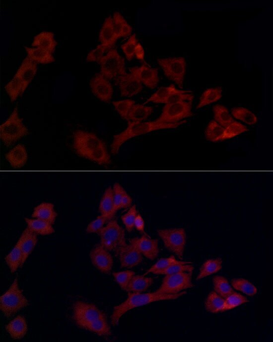
- Immunofluorescence analysis of UBE2C in NIH/3T3 cells. Samples were incubated with UBE2C Polyclonal antibody (Product # PA5-117019) using a dilution of 1:100 (40x lens). Blue: DAPI for nuclear staining.
- Submitted by
- Invitrogen Antibodies (provider)
- Main image
- Experimental details
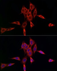
- Immunofluorescence analysis of UBE2C in PC-12 cells. Samples were incubated with UBE2C Polyclonal antibody (Product # PA5-117019) using a dilution of 1:100 (40x lens). Blue: DAPI for nuclear staining.
Supportive validation
- Submitted by
- Invitrogen Antibodies (provider)
- Main image

- Experimental details
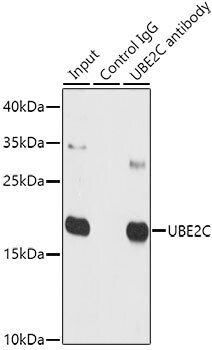
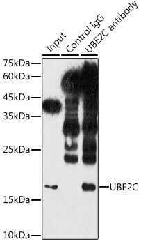
- Immunoprecipitation of UBE2C in 300 μg extracts of NIH/3T3 cells. Samples were precipitated with 3 μg UBE2C Polyclonal antibody (Product # PA5-117019). Western blot was performed from the immunoprecipitate using UBE2C Polyclonal antibody (Product # PA5-117019) at a dilution of 1:1,000.
Supportive validation
- Submitted by
- Invitrogen Antibodies (provider)
- Main image
- Experimental details
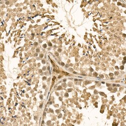
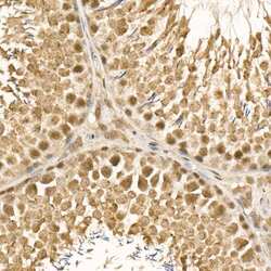
- Immunohistochemistry analysis of UBE2C in paraffin-embedded mouse testis. Samples were incubated with UBE2C Polyclonal antibody (Product # PA5-117019) using a dilution of 1:100 (40x lens). Perform high pressure antigen retrieval with 10 mM citrate buffer pH 6.0 before commencing with IHC staining protocol.
- Submitted by
- Invitrogen Antibodies (provider)
- Main image
- Experimental details
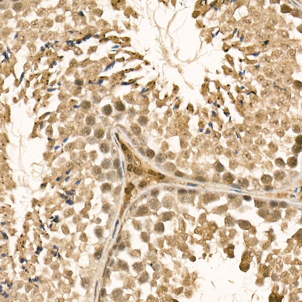
- Immunohistochemistry analysis of UBE2C in paraffin-embedded rat testis. Samples were incubated with UBE2C Polyclonal antibody (Product # PA5-117019) using a dilution of 1:100 (40x lens). Perform high pressure antigen retrieval with 10 mM citrate buffer pH 6.0 before commencing with IHC staining protocol.

Supportive validation
- Submitted by
- Invitrogen Antibodies (provider)
- Main image

- Experimental details
- Immunoprecipitation analysis of UBE2C in 150 µg extracts of SW620 cells using UBE2C Polyclonal Antibody (Product # PA5-117019). Western blot was performed from the immunoprecipitate using the same antibody at a dilution of 1:500