Explore
 Validate
 Learn
Learn40-1500
antibody from Invitrogen Antibodies
Targeting: FBXW7
AGO, CDC4, FBW7, FBX30, FBXW6, FLJ11071, SEL-10, SEL10
 Western blot
 Immunoprecipitation
 Immunohistochemistry
Antibody data
- Antibody Data
- Antigen structure
- References [6]
- Comments [0]
- Validations
- Immunohistochemistry [3]
- Flow cytometry [1]
- Other assay [3]
Submit
Validation data
Reference
Comment
Report error
- Product number
- 40-1500 - Provider product page

- Provider
- Invitrogen Antibodies
- Product name
- FBXW7 Polyclonal Antibody
- Antibody type
- Polyclonal
- Antigen
- Synthetic peptide
- Reactivity
- Human, Mouse
- Host
- Rabbit
- Isotype
- IgG
- Vial size
- 100 μg
- Concentration
- 0.25 mg/mL
- Storage
- -20°C
Submitted references Tanshinone IIA inhibits oral squamous cell carcinoma via reducing Akt-c-Myc signaling-mediated aerobic glycolysis.
Fbxw7 increases CCL2/7 in CX3CR1hi macrophages to promote intestinal inflammation.
Ubiquitin ligase RNF8 suppresses Notch signaling to regulate mammary development and tumorigenesis.
PLK1 stabilizes a MYC-dependent kinase network in aggressive B cell lymphomas.
E3 ligase FBXW7 is critical for RIG-I stabilization during antiviral responses.
Polo-like Kinase-1 Regulates Myc Stabilization and Activates a Feedforward Circuit Promoting Tumor Cell Survival.
Li M, Gao F, Zhao Q, Zuo H, Liu W, Li W
Cell death & disease 2020 May 18;11(5):381
Cell death & disease 2020 May 18;11(5):381
Fbxw7 increases CCL2/7 in CX3CR1hi macrophages to promote intestinal inflammation.
He J, Song Y, Li G, Xiao P, Liu Y, Xue Y, Cao Q, Tu X, Pan T, Jiang Z, Cao X, Lai L, Wang Q
The Journal of clinical investigation 2019 Jun 27;129(9):3877-3893
The Journal of clinical investigation 2019 Jun 27;129(9):3877-3893
Ubiquitin ligase RNF8 suppresses Notch signaling to regulate mammary development and tumorigenesis.
Li L, Guturi KKN, Gautreau B, Patel PS, Saad A, Morii M, Mateo F, Palomero L, Barbour H, Gomez A, Ng D, Kotlyar M, Pastrello C, Jackson HW, Khokha R, Jurisica I, Affar EB, Raught B, Sanchez O, Alaoui-Jamali M, Pujana MA, Hakem A, Hakem R
The Journal of clinical investigation 2018 Oct 1;128(10):4525-4542
The Journal of clinical investigation 2018 Oct 1;128(10):4525-4542
PLK1 stabilizes a MYC-dependent kinase network in aggressive B cell lymphomas.
Ren Y, Bi C, Zhao X, Lwin T, Wang C, Yuan J, Silva AS, Shah BD, Fang B, Li T, Koomen JM, Jiang H, Chavez JC, Pham LV, Sudalagunta PR, Wan L, Wang X, Dalton WS, Moscinski LC, Shain KH, Vose J, Cleveland JL, Sotomayor EM, Fu K, Tao J
The Journal of clinical investigation 2018 Dec 3;128(12):5517-5530
The Journal of clinical investigation 2018 Dec 3;128(12):5517-5530
E3 ligase FBXW7 is critical for RIG-I stabilization during antiviral responses.
Song Y, Lai L, Chong Z, He J, Zhang Y, Xue Y, Xie Y, Chen S, Dong P, Chen L, Chen Z, Dai F, Wan X, Xiao P, Cao X, Liu Y, Wang Q
Nature communications 2017 Mar 13;8:14654
Nature communications 2017 Mar 13;8:14654
Polo-like Kinase-1 Regulates Myc Stabilization and Activates a Feedforward Circuit Promoting Tumor Cell Survival.
Xiao D, Yue M, Su H, Ren P, Jiang J, Li F, Hu Y, Du H, Liu H, Qing G
Molecular cell 2016 Nov 3;64(3):493-506
Molecular cell 2016 Nov 3;64(3):493-506
No comments: Submit comment
Supportive validation
- Submitted by
- Invitrogen Antibodies (provider)
- Main image
- Experimental details
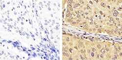
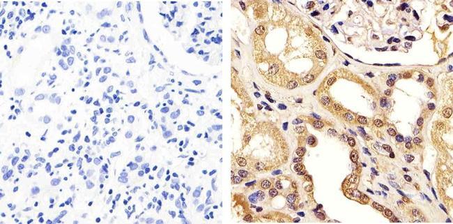
- Immunohistochemistry analysis of Cdc4 showing staining in the cytoplasm and nucleus of paraffin-embedded human cervical carcinoma tissue (right) compared to a negative control without primary antibody (left). To expose target proteins, antigen retrieval was performed using 10mM sodium citrate (pH 6.0), microwaved for 8-15 min. Following antigen retrieval, tissues were blocked in 3% H2O2-methanol for 15 min at room temperature, washed with ddH2O and PBS, and then probed with a Cdc4 polyclonal antibody (Product # 40-1500) diluted in 3% BSA-PBS at a dilution of 1:50 overnight at 4ºC in a humidified chamber. Tissues were washed extensively in PBST and detection was performed using an HRP-conjugated secondary antibody followed by colorimetric detection using a DAB kit. Tissues were counterstained with hematoxylin and dehydrated with ethanol and xylene to prep for mounting.
- Submitted by
- Invitrogen Antibodies (provider)
- Main image
- Experimental details
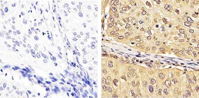
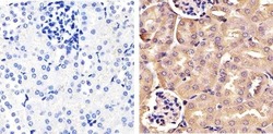
- Immunohistochemistry analysis of Cdc4 showing staining in the cytoplasm and nucleus of paraffin-embedded human kidney tissue (right) compared to a negative control without primary antibody (left). To expose target proteins, antigen retrieval was performed using 10mM sodium citrate (pH 6.0), microwaved for 8-15 min. Following antigen retrieval, tissues were blocked in 3% H2O2-methanol for 15 min at room temperature, washed with ddH2O and PBS, and then probed with a Cdc4 polyclonal antibody (Product # 40-1500) diluted in 3% BSA-PBS at a dilution of 1:50 overnight at 4ºC in a humidified chamber. Tissues were washed extensively in PBST and detection was performed using an HRP-conjugated secondary antibody followed by colorimetric detection using a DAB kit. Tissues were counterstained with hematoxylin and dehydrated with ethanol and xylene to prep for mounting.
- Submitted by
- Invitrogen Antibodies (provider)
- Main image
- Experimental details
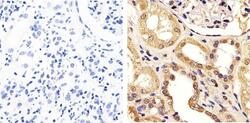
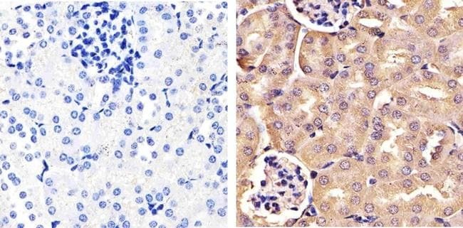
- Immunohistochemistry analysis of Cdc4 showing staining in the cytoplasm and nucleus of paraffin-embedded mouse kidney tissue (right) compared to a negative control without primary antibody (left). To expose target proteins, antigen retrieval was performed using 10mM sodium citrate (pH 6.0), microwaved for 8-15 min. Following antigen retrieval, tissues were blocked in 3% H2O2-methanol for 15 min at room temperature, washed with ddH2O and PBS, and then probed with a Cdc4 polyclonal antibody (Product # 40-1500) diluted in 3% BSA-PBS at a dilution of 1:20 overnight at 4ºC in a humidified chamber. Tissues were washed extensively in PBST and detection was performed using an HRP-conjugated secondary antibody followed by colorimetric detection using a DAB kit. Tissues were counterstained with hematoxylin and dehydrated with ethanol and xylene to prep for mounting.
Supportive validation
- Submitted by
- Invitrogen Antibodies (provider)
- Main image
- Experimental details
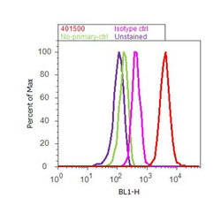
- Flow cytometry analysis of Cdc4 was done on HeLa. Cells were fixed with 70% ethanol for 10 minutes, permeabilized with 0.25% Triton™ X-100 for 20 minutes, and blocked with 5% BSA for 30 minutes at room temperature. Cells were labeled with Cdc4 Rabbit Polyclonal Antibody (401500, red histogram) or with rabbit isotype control (pink histogram) at 3-5 ug/million cells in 2.5% BSA. After incubation at room temperature for 2 hours, the cells were labeled with Alexa Fluor® 488 Goat Anti-Rabbit Secondary Antibody (A11008) at a dilution of 1:400 for 30 minutes at room temperature. The representative 10,000 cells were acquired and analyzed for each sample using an Attune® Acoustic Focusing Cytometer. The purple histogram represents unstained control cells and the green histogram represents no-primary-antibody control.

Supportive validation
- Submitted by
- Invitrogen Antibodies (provider)
- Main image
- Experimental details
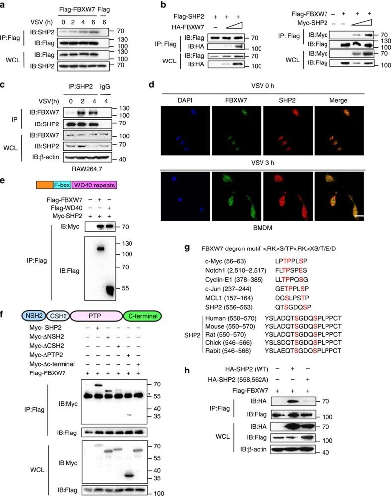
- Figure 5 FBXW7 interacts with SHP2. ( a ) Coimmunoprecipitation and immunoblot analysis of HEK293T cells transfected for 24 h with Flag-FBXW7 cells infected with VSV and treated with MG132. ( b ) Coimmunoprecipitation and immunoblot analysis of HEK293T cells cotransfected for 24 h with Flag-SHP2 plus HA-FBXW7 or Flag-FBXW7 and Myc-SHP2, and treated with MG132 followed by immunoprecipitation with anti-Flag-M2 beads. ( c ) Immunoblot analysis of RAW264.7 infected for indicate hours with VSV and treated with MG132, followed by immunoprecipitation with SHP2-conjugated agarose or immunoglobulin G (IgG)-conjugated agarose. ( d ) Confocal microscopy imaging of peritoneal macrophages infected for 3 h with VSV and labelled with antibodies to the appropriate protein. Scale bar, 20 mum. ( e ) Immunoblot analysis of HEK293T cells cotransfected for 24 h with Flag-FBXW7 or Flag-WD40, plus Myc-SHP2 vectors and treated with MG132, followed by immunoprecipitation with anti-Flag-M2 beads. ( f ) Immunoblot analysis of HEK293T cells cotransfected for 48 h with Flag-FBXW7, plus Myc-SHP2, Myc-SHP2 mutants vectors and treated with MG132, followed by immunoprecipitation with anti-Flag-M2 beads. *IgG heavy chain. ( g ) Sequence alignment of SHP2 with FBXW7 degron motif ( S/TP XS/T/E/D), where X is any amino acid and is any amino acid, except arginine (R) or lysine (K). ( h ) Coimmunoprecipitation and immunoblot analysis of HEK293T cells transfected with Flag-FBXW7 along with vector for H
- Submitted by
- Invitrogen Antibodies (provider)
- Main image
- Experimental details
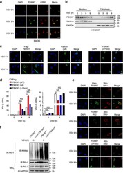
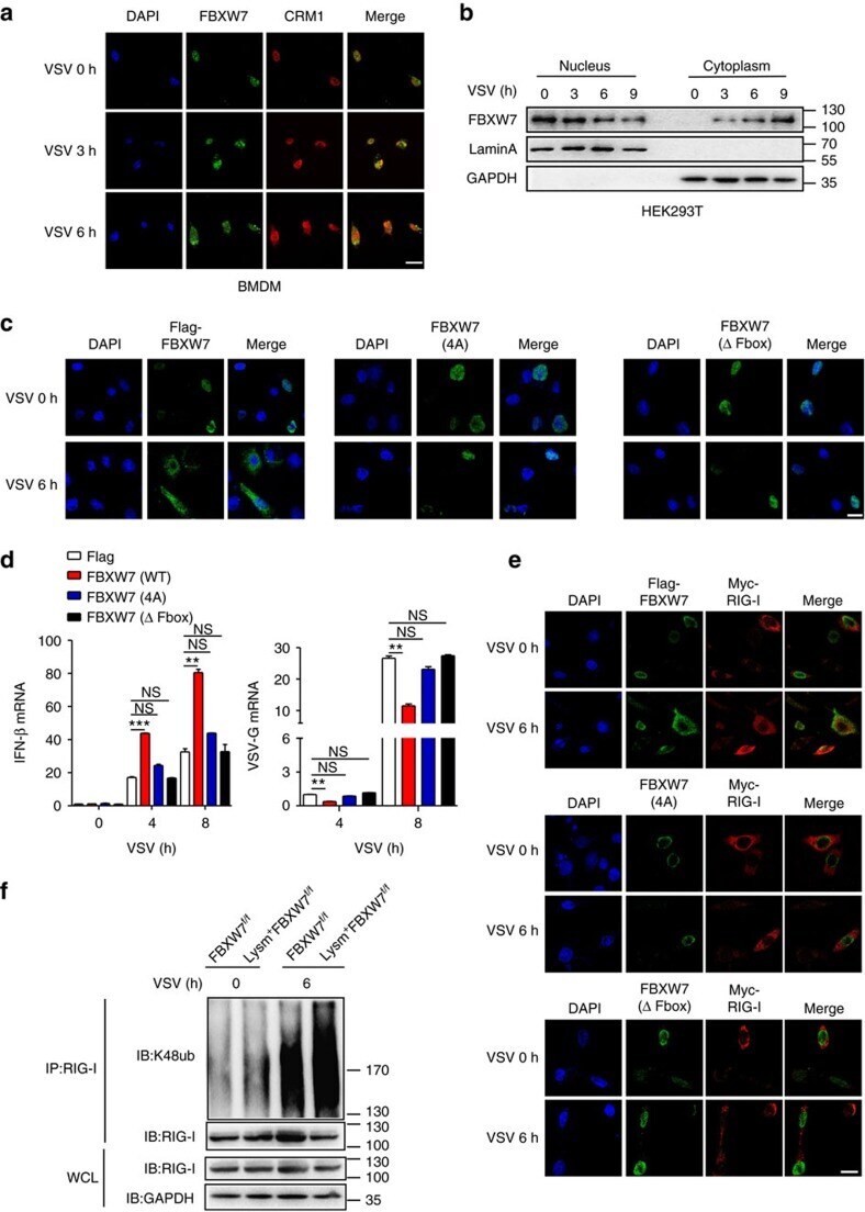
- Figure 4 FBXW7 translocates into the cytoplasm to interact with RIG-I and exert the antiviral function. ( a ) Confocal microscopy imaging of BMDM infected for indicated hours with VSV and labelled with antibodies to the appropriate protein. Scale bar, 20 mum. ( b ) Immunoblot analysis of Flag-FBXW7 among nuclear and cytoplasm proteins from overexpressed Flag-FBXW7 HEK293T cells infected with VSV; Lamin A serves as a nuclear control; GAPDH serves as a cytoplasm control. ( c ) Confocal microscopy imaging of HEK293T cells transfected with Flag-FBXW7 (wt) or Flag-FBXW7 (mutant) plasmid for 24 h, then infected for indicated hours with VSV and labelled with antibodies to the appropriate protein. Scale bar, 20 mum. ( d ) Quantitative PCR (Q-PCR) analysis of IFN-beta and VSV-G mRNA expression in HEK293T cells transfected with Flag-FBXW7 (wt) or Flag-FBXW7 (mutant) plasmid and infected with VSV for indicated hours. ( e ) Confocal microscopy imaging of HEK293T cells that transfected with Flag-FBXW7 (wt) or Flag-FBXW7 (mutant) together with Myc-RIG-I plasmids followed by VSV infection for 6 h. Scale bar, 20 mum. ( f ) Immunoblot analysis of the K48 ubiquitination of RIG-I in FBXW7 f/f and Lysm + FBXW7 f/f peritoneal macrophages infected with VSV. Data are mean+-s.e.m. and are representative of three independent experiments. Student's t -test was used for statistical calculation. ** P
- Submitted by
- Invitrogen Antibodies (provider)
- Main image

- Experimental details
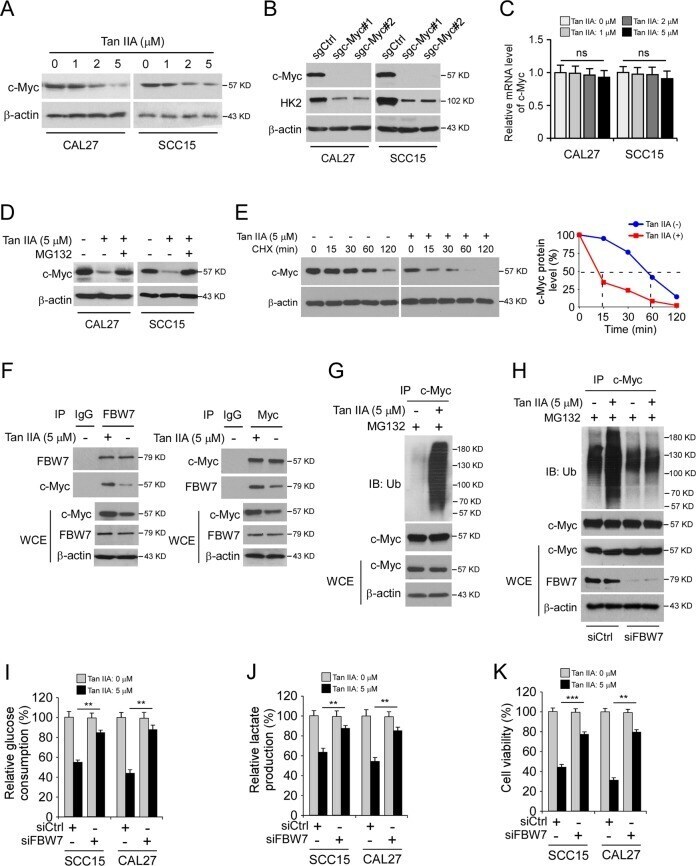
- Fig. 4 Tan IIA promotes c-Myc ubiquitination and degradation. a IB analysis of c-Myc expression in OSCC cells with Tan IIA treatment. b IB analysis of c-Myc and HK2 expression in sgCtrl and sgc-Myc expressing OSCC cells. c qRT-PCR analysis of c-Myc mRNA level in Tan IIA-treated OSCC cells. ns, not statistically significant. d OSCC cells were treated with Tan IIA for 24 h, MG132 was added to the cell culture medium and incubated for another 6 h, whole-cell lysate (WCE) was subjected to IB analysis. e OSCC cells were treated with Tan IIA for 24 h, cycloheximide (CHX) was added to the cell culture medium and incubated for various time points as indicated, whole-cell lysate (WCE) was subjected to IB analysis. f co-IP analysis of the interaction between c-Myc and FBW7 in Tan IIA-treated CAL27 cells. g Ubiquitination analysis of c-Myc ubiquitination in Tan IIA-treated CAL27 cells. h CAL27 cells were transfected with siCtrl or siFBW7 shRNA followed by Tan IIA-treated for 24 h, WCE was subjected to ubiquitination analysis. i , j CAL27 cells were transfected with siCtrl or siFBW7 shRNA followed by Tan IIA-treated for 24 h, the cell culture medium was subjected to glucose consumption ( i ) and lactate production ( j ) analysis. k CAL27 cells were transfected with siCtrl or siFBW7 shRNA followed by Tan IIA-treated for 24 h and subjected to MTS assay for cell viability analysis. ** p < 0.01, *** p < 0.001.