Explore
 Validate
 Learn
Learn Western blot
 Immunocytochemistry
Antibody data
- Antibody Data
- Antigen structure
- References [1]
- Comments [0]
- Validations
- Immunocytochemistry [1]
- Other assay [1]
Submit
Validation data
Reference
Comment
Report error
- Product number
- 702444 - Provider product page

- Provider
- Invitrogen Antibodies
- Product name
- PIP5K1C Recombinant Rabbit Monoclonal Antibody (16H4L5)
- Antibody type
- Monoclonal
- Antigen
- Synthetic peptide
- Description
- This antibody is predicted to react with Monkey, Pig, Mouse, Bovine Recombinant rabbit monoclonal antibodies are produced using in vitro expression systems. The expression systems are developed by cloning in the specific antibody DNA sequences from immunoreactive rabbits. Then, individual clones are screened to select the best candidates for production. The advantages of using recombinant rabbit monoclonal antibodies include: better specificity and sensitivity, lot-to-lot consistency, animal origin-free formulations, and broader immunoreactivity to diverse targets due to larger rabbit immune repertoire.
- Reactivity
- Human
- Host
- Rabbit
- Isotype
- IgG
- Antibody clone number
- 16H4L5
- Vial size
- 100 μg
- Concentration
- 0.5 mg/mL
- Storage
- Store at 4°C short term. For long term storage, store at -20°C, avoiding freeze/thaw cycles.
Submitted references Lipid Messenger Phosphatidylinositol-4,5-Bisphosphate Is Increased by Both PPARα Activators and Inhibitors: Relevance for Intestinal Cell Differentiation.
Cizkova K, Koubova K, Tauber Z
Biology 2022 Jun 30;11(7)
Biology 2022 Jun 30;11(7)
No comments: Submit comment
Supportive validation
- Submitted by
- Invitrogen Antibodies (provider)
- Main image
- Experimental details
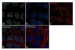
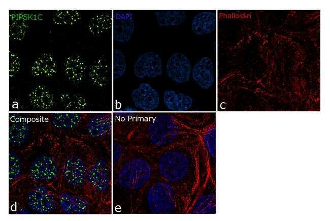
- For immunofluorescence analysis, A431 cells were fixed and permeabilized for detection of endogenous Phosphatidylinositol 4-phosphate 5-kinase type-1 gamma using Anti- PIP5K1C Recombinant Rabbit Monoclonal Antibody (Product # 702444, 5 µg/mL) and labeled with Goat anti-Rabbit IgG (Heavy Chain) Superclonal™ Secondary Antibody, Alexa Fluor® 488 conjugate (Product # A27034, 1:2000). Panel a) shows representative cells that were stained for detection and localization of Phosphatidylinositol 4-phosphate 5-kinase type-1 gamma protein (green), Panel b) is stained for nuclei (blue) using SlowFade® Gold Antifade Mountant with DAPI (Product # S36938). Panel c) represents cytoskeletal F-actin staining using Rhodamine Phalloidin (Product # R415, 1:300). Panel d) is a composite image of Panels a, b and c clearly demonstrating nuclear localisation of PIP5K1C. Panel e) represents control cells with no primary antibody to assess background. The images were captured at 60X magnification.
Supportive validation
- Submitted by
- Invitrogen Antibodies (provider)
- Main image
- Experimental details
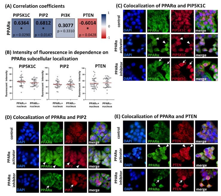
- Relationships between levels of PPARalpha and PIP5K1C, PIP2, PI3K, and PTEN in HT-29 cell line. ( A ) Spearman correlation coefficients ( n = 12) for expression levels obtained with In-Cell ELISA method. PPARalpha expression under the same experimental condition was examined in our previous study []. Moderate positive correlations were found between PPARalpha and PIP5K1C and PPARalpha and PIP2, whereas PPARalpha and PTEN expression showed moderate negative correlation (statistical significance marked with asterisk, * p