 Explore
 Validate
 Learn
LearnMA1-038
antibody from Invitrogen Antibodies
Targeting: OGT
FLJ23071, HRNT1, MGC22921, O-GLCNAC, OGT1
 Western blot
 Immunoprecipitation
Antibody data
- Antibody Data
- Antigen structure
- References [2]
- Comments [0]
- Validations
- Immunoprecipitation [1]
- Other assay [1]
Submit
Validation data
Reference
Comment
Report error
- Product number
- MA1-038 - Provider product page

- Provider
- Invitrogen Antibodies
- Product name
- Anti-O-linked N-acetylglucosamine (O-GlcNAc) Monoclonal Antibody (18B10.C7)
- Antibody type
- Monoclonal
- Antigen
- Synthetic peptide
- Description
- MA1-038 has been successfully used in Western blot and immunoprecipitation applications with human samples. In Western blot, MA1-038 detects proteins with O-GlcNAc post-translational modification, and therefore will detect multiple specific bands.
- Reactivity
- Human
- Host
- Mouse
- Isotype
- IgG
- Antibody clone number
- 18B10.C7
- Vial size
- 100 µg
- Concentration
- 1 mg/mL
- Storage
- -20° C, Avoid Freeze/Thaw Cycles
Submitted references O-GlcNAcylation of G6PD promotes the pentose phosphate pathway and tumor growth.
The intracellular fate of zonula occludens 2 is regulated by the phosphorylation of SR repeats and the phosphorylation/O-GlcNAcylation of S257.
Rao X, Duan X, Mao W, Li X, Li Z, Li Q, Zheng Z, Xu H, Chen M, Wang PG, Wang Y, Shen B, Yi W
Nature communications 2015 Sep 24;6:8468
Nature communications 2015 Sep 24;6:8468
The intracellular fate of zonula occludens 2 is regulated by the phosphorylation of SR repeats and the phosphorylation/O-GlcNAcylation of S257.
Quiros M, Alarcón L, Ponce A, Giannakouros T, González-Mariscal L
Molecular biology of the cell 2013 Aug;24(16):2528-43
Molecular biology of the cell 2013 Aug;24(16):2528-43
No comments: Submit comment
Supportive validation
- Submitted by
- Invitrogen Antibodies (provider)
- Main image

- Experimental details
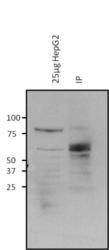
- Immunoprecipitation of Glycogen Synthase was performed on HepG2 cells, glucose and serum starved for 36 hours. The antigen:antibody complex was formed by binding 500 µg whole cell lysate with 5 µg of mouse monoclonal antibody recognizing O-GlcNAc (Product # MA1-038) overnight on a rocking platform at 4°C. The immune-complex was then captured on 50 µL Protein A/G Plus Agarose (Product # 20424). The beads were washed to remove non-bound material and a low-pH elution buffer was used to dissociate bound antigen from the antibody-bound bead. Samples were then resolved on a 4-20% Tris-HCl polyacrylamide gel. Proteins were transferred to PVDF membrane and blocked with 5% BSA/TBS-0.1%Tween for at least 1 hour. Membranes were then probed with a mouse monoclonal antibody recognizing Glycogen Synthase (Product # PA5-17459) at a dilution of 1:1000 overnight rotating at 4°C. Membranes were then washed in TBST and probed with Clean-blot IP detection reagent (Product # 21230) at a dilution of 1:2000 for at least one hour. Membranes were washed and chemiluminescent detection was performed using Super Signal West Pico (Product # 34087). The image is a Western blot with antibody Product # PA5-17459 showing enrichment of glycogen synthase by O-GlcNAc precipitation. Lane 1 indicates 25 µg of HepG2 whole cell lysate input; Lane 2 indicates the IP elution.

Supportive validation
- Submitted by
- Invitrogen Antibodies (provider)
- Main image
- Experimental details
- Immunoprecipitation of Glycogen Synthase was performed on HepG2 cells, glucose and serum starved for 36 hours. The antigen:antibody complex was formed by binding 500 µg whole cell lysate with 5 µg of mouse monoclonal antibody recognizing O-GlcNAc (Product # MA1-038) overnight on a rocking platform at 4°C. The immune-complex was then captured on 50 µL Protein A/G Plus Agarose (Product # 20424). The beads were washed to remove non-bound material and a low-pH elution buffer was used to dissociate bound antigen from the antibody-bound bead. Samples were then resolved on a 4-20% Tris-HCl polyacrylamide gel. Proteins were transferred to PVDF membrane and blocked with 5% BSA/TBS-0.1%Tween for at least 1 hour. Membranes were then probed with a mouse monoclonal antibody recognizing Glycogen Synthase (Product # PA5-17459) at a dilution of 1:1000 overnight rotating at 4°C. Membranes were then washed in TBST and probed with Clean-blot IP detection reagent (Product # 21230) at a dilution of 1:2000 for at least one hour. Membranes were washed and chemiluminescent detection was performed using Super Signal West Pico (Product # 34087). The image is a Western blot with antibody Product # PA5-17459 showing enrichment of glycogen synthase by O-GlcNAc precipitation. Lane 1 indicates 25 µg of HepG2 whole cell lysate input; Lane 2 indicates the IP elution.
