 Explore
 Validate
 Learn
LearnPA5-22071
antibody from Invitrogen Antibodies
Targeting: OGT
FLJ23071, HRNT1, MGC22921, O-GLCNAC, OGT1
 Western blot
 Immunocytochemistry Immunoprecipitation Immunohistochemistry
 Flow cytometry Other assay
Antibody data
- Antibody Data
- Antigen structure
- References [2]
- Comments [0]
- Validations
- Immunocytochemistry [6]
- Immunoprecipitation [1]
- Immunohistochemistry [6]
- Other assay [2]
Submit
Validation data
Reference
Comment
Report error
- Product number
- PA5-22071 - Provider product page

- Provider
- Invitrogen Antibodies
- Product name
- OGT Polyclonal Antibody
- Antibody type
- Polyclonal
- Antigen
- Recombinant full-length protein
- Description
- Recommended positive controls: 293T, A431, HeLa, HepG2, mouse brain, PC-12. Predicted reactivity: Mouse (100%), Rat (100%), Xenopus laevis (98%), Pig (100%), Bovine (100%). Store product as a concentrated solution. Centrifuge briefly prior to opening the vial.
- Reactivity
- Human, Mouse, Rat
- Host
- Rabbit
- Isotype
- IgG
- Vial size
- 100 μL
- Concentration
- 1.51 mg/mL
- Storage
- Store at 4°C short term. For long term storage, store at -20°C, avoiding freeze/thaw cycles.
Submitted references O-GlcNAc transferase regulates p21 protein levels and cell proliferation through the FoxM1-Skp2 axis in a p53-independent manner.
Redirected nuclear glutamate dehydrogenase supplies Tet3 with α-ketoglutarate in neurons.
de Queiroz RM, Moon SH, Prives C
The Journal of biological chemistry 2022 Sep;298(9):102289
The Journal of biological chemistry 2022 Sep;298(9):102289
Redirected nuclear glutamate dehydrogenase supplies Tet3 with α-ketoglutarate in neurons.
Traube FR, Özdemir D, Sahin H, Scheel C, Glück AF, Geserich AS, Oganesian S, Kostidis S, Iwan K, Rahimoff R, Giorgio G, Müller M, Spada F, Biel M, Cox J, Giera M, Michalakis S, Carell T
Nature communications 2021 Jul 2;12(1):4100
Nature communications 2021 Jul 2;12(1):4100
No comments: Submit comment
Supportive validation
- Submitted by
- Invitrogen Antibodies (provider)
- Main image

- Experimental details
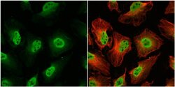
- Immunocytochemistry-Immunofluorescence analysis of OGT was performed in HeLa cells fixed in 4% paraformaldehyde at RT for 15 min. Green: OGT Polyclonal Antibody (Product # PA5-22071) diluted at 1:200. Red: alpha Tubulin, a cytoskeleton marker.

- Submitted by
- Invitrogen Antibodies (provider)
- Main image

- Experimental details
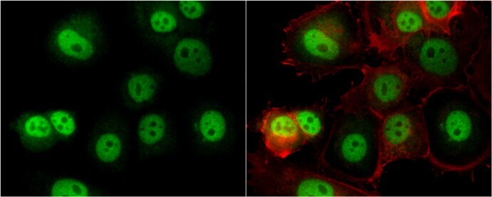
- Immunocytochemistry-Immunofluorescence analysis of OGT was performed in MCF7 cells fixed in 4% paraformaldehyde at RT for 15 min. Green: OGT Polyclonal Antibody (Product # PA5-22071) diluted at 1:500. Red: phalloidin, a cytoskeleton marker.

- Submitted by
- Invitrogen Antibodies (provider)
- Main image

- Experimental details
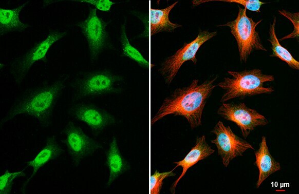
- OGT Polyclonal Antibody detects O-GlcNAc transferase protein at nucleus by immunofluorescent analysis. Sample: HeLa cells were fixed in 4% paraformaldehyde at RT for 15 min. Green: O-GlcNAc transferase stained by OGT Polyclonal Antibody (Product # PA5-22071) diluted at 1:1,000. Red: alpha Tubulin, a cytoskeleton marker, stained by alpha Tubulin Polyclonal Antibody [GT114] (Product # MA5-31466) diluted at 1:1,000. Blue: Fluoroshield with DAPI .

- Submitted by
- Invitrogen Antibodies (provider)
- Main image

- Experimental details
- OGT Polyclonal Antibody detects O-GlcNAc transferase protein at nucleus by immunofluorescent analysis. Sample: HeLa cells were fixed in 4% paraformaldehyde at RT for 15 min. Green: O-GlcNAc transferase stained by OGT Polyclonal Antibody (Product # PA5-22071) diluted at 1:1,000. Red: alpha Tubulin, a cytoskeleton marker, stained by alpha Tubulin Polyclonal Antibody [GT114] (Product # MA5-31466) diluted at 1:1,000. Blue: Fluoroshield with DAPI .
- Submitted by
- Invitrogen Antibodies (provider)
- Main image
- Experimental details
- Immunocytochemistry-Immunofluorescence analysis of OGT was performed in HeLa cells fixed in 4% paraformaldehyde at RT for 15 min. Green: OGT Polyclonal Antibody (Product # PA5-22071) diluted at 1:200. Red: alpha Tubulin, a cytoskeleton marker.

- Submitted by
- Invitrogen Antibodies (provider)
- Main image

- Experimental details
- Immunocytochemistry-Immunofluorescence analysis of OGT was performed in MCF7 cells fixed in 4% paraformaldehyde at RT for 15 min. Green: OGT Polyclonal Antibody (Product # PA5-22071) diluted at 1:500. Red: phalloidin, a cytoskeleton marker.
Supportive validation
- Submitted by
- Invitrogen Antibodies (provider)
- Main image

- Experimental details
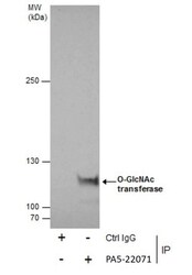
- Immunoprecipitation of O-GlcNAc transferase was performed in A431 whole cell extracts using 5 µg of OGT Polyclonal Antibody (Product # PA5-22071). Samples were transferred to a membrane and probed with OGT Polyclonal Antibody as a primary antibody and an HRP-conjugated anti-Rabbit IgG was used as a secondary antibody.

Supportive validation
- Submitted by
- Invitrogen Antibodies (provider)
- Main image
- Experimental details
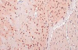
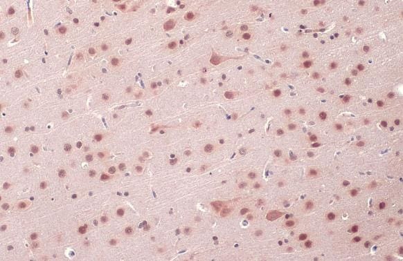
- OGT Polyclonal Antibody detects O-GlcNAc transferase protein at cytoplasm and nucleus by immunohistochemical analysis. Sample: Paraffin-embedded mouse brain. O-GlcNAc transferase stained by OGT Polyclonal Antibody (Product # PA5-22071) diluted at 1:500. Antigen Retrieval: Citrate buffer, pH 6.0, 15 min.
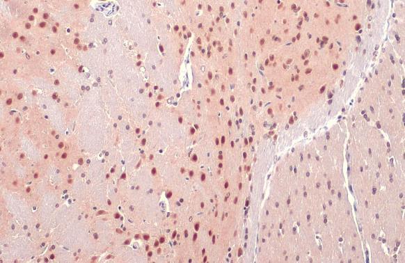
- Submitted by
- Invitrogen Antibodies (provider)
- Main image
- Experimental details
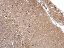
- OGT Polyclonal Antibody detects O-GlcNAc transferase protein at cytoplasm and nucleus by immunohistochemical analysis. Sample: Paraffin-embedded mouse brain. O-GlcNAc transferase stained by OGT Polyclonal Antibody (Product # PA5-22071) diluted at 1:500. Antigen Retrieval: Citrate buffer, pH 6.0, 15 min.
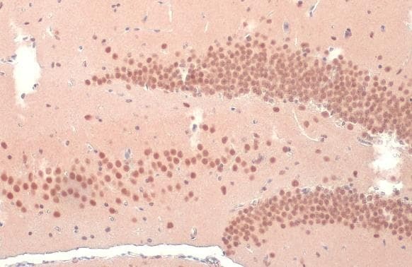
- Submitted by
- Invitrogen Antibodies (provider)
- Main image

- Experimental details
- OGT Polyclonal Antibody detects O-GlcNAc transferase protein at cytoplasm and nucleus by immunohistochemical analysis. Sample: Paraffin-embedded mouse brain. O-GlcNAc transferase stained by OGT Polyclonal Antibody (Product # PA5-22071) diluted at 1:500. Antigen Retrieval: Citrate buffer, pH 6.0, 15 min.
- Submitted by
- Invitrogen Antibodies (provider)
- Main image

- Experimental details
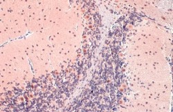
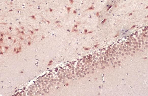
- OGT Polyclonal Antibody detects O-GlcNAc transferase protein at cytoplasm and nucleus by immunohistochemical analysis. Sample: Paraffin-embedded rat brain. O-GlcNAc transferase stained by OGT Polyclonal Antibody (Product # PA5-22071) diluted at 1:500. Antigen Retrieval: Citrate buffer, pH 6.0, 15 min.
- Submitted by
- Invitrogen Antibodies (provider)
- Main image
- Experimental details
- OGT Polyclonal Antibody detects O-GlcNAc transferase protein at cytoplasm and nucleus by immunohistochemical analysis. Sample: Paraffin-embedded rat brain. O-GlcNAc transferase stained by OGT Polyclonal Antibody (Product # PA5-22071) diluted at 1:500. Antigen Retrieval: Citrate buffer, pH 6.0, 15 min.
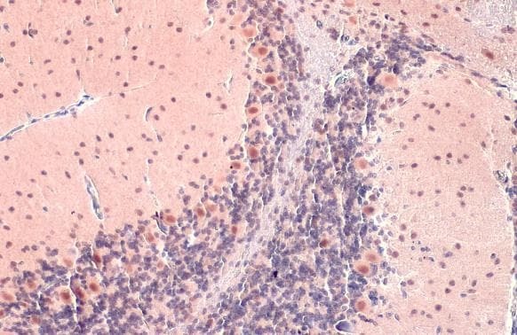
- Submitted by
- Invitrogen Antibodies (provider)
- Main image
- Experimental details
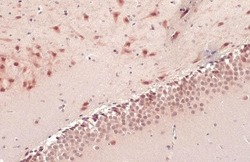
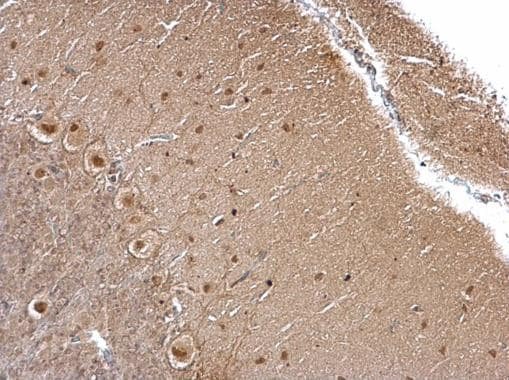
- OGT Polyclonal Antibody detects O-GlcNAc transferase protein at cytoplasm and nucleus on rat hind brain by immunohistochemical analysis. Sample: Paraffin-embedded rat hind brain. OGT Polyclonal Antibody (Product # PA5-22071) diluted at 1:500. Antigen Retrieval: EDTA based buffer, pH 8.0, 15 min.
Supportive validation
- Submitted by
- Invitrogen Antibodies (provider)
- Main image
- Experimental details
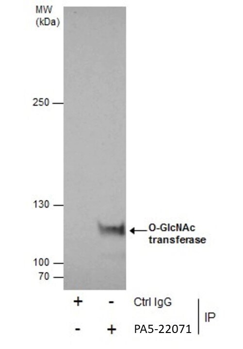
- Immunoprecipitation of O-GlcNAc transferase was performed in A431 whole cell extracts using 5 µg of OGT Polyclonal Antibody (Product # PA5-22071). Samples were transferred to a membrane and probed with OGT Polyclonal Antibody as a primary antibody and an HRP-conjugated anti-Rabbit IgG was used as a secondary antibody.
- Submitted by
- Invitrogen Antibodies (provider)
- Main image
- Experimental details
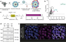
- Fig. 2 Interaction of Tet3 with Gdh in neurons. a Experimental set-up for Tet3-enriched coIP. b Volcano plot after Tet3-enriched coIP in nuclear brain lysate of adult mice ( n = 4 biologically independent sample preparations). Interaction partners were analyzed using label-free quantification after LC-MS/MS. Enriched interaction partners (FDR < 0.05, log 2 FC > 1) are highlighted in black. c Conversion of glutamate to alphaKG by Gdh and glutamate to alphaKG ratio determined by NMR-based metabolomics in murine hippocampus ( n = 6 biologically independent animals, bar shows mean, error bars SD). d Western blots detecting Tet3, Ogt, and Gdh after endogenous Tet3-coIP in nuclear brain lysate. e PLA signal (white/orange dots) of Tet3 and Gdh in the dentate gyrus (DG) of the murine hippocampus. Nuclei (blue) were stained with Hoechst and NeuN staining indicates neuronal nuclei. Scale bar is 5 um. Source data are provided as a Source Data file.
