Explore
 Validate
 Learn
Learn Western blot
 Immunocytochemistry
 Immunoprecipitation
Antibody data
- Antibody Data
- Antigen structure
- References [0]
- Comments [0]
- Validations
- Western blot [2]
- Immunoprecipitation [3]
- Immunohistochemistry [16]
Submit
Validation data
Reference
Comment
Report error
- Product number
- LS-C784225 - Provider product page

- Provider
- LSBio
- Product name
- CHEK2 / CHK2 Antibody (clone OTI5C4, Carrier-free) LS-C784225
- Antibody type
- Monoclonal
- Description
- Purified from ascites.
- Reactivity
- Human, Canine
- Host
- Mouse
- Isotype
- IgG
- Antibody clone number
- OTI5C4
- Storage
- Store at -20°C. Avoid freeze-thaw cycles.
No comments: Submit comment
Enhanced validation
- Submitted by
- LSBio (provider)
- Enhanced method
- Genetic validation
- Main image
- Experimental details
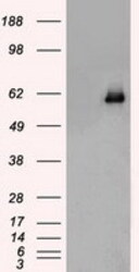
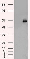
- HEK293T cells were transfected with the pCMV6-ENTRY control (Left lane) or pCMV6-ENTRY CHEK2 (Right lane) cDNA for 48 hrs and lysed. Equivalent amounts of cell lysates (5 ug per lane) were separated by SDS-PAGE and immunoblotted with anti-CHEK2.
- Submitted by
- LSBio (provider)
- Enhanced method
- Genetic validation
- Main image
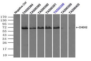
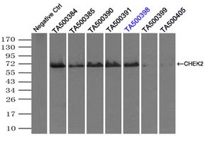
- Experimental details
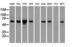
- Western blot of extracts (35 ug) from 9 different cell lines by using anti-CHEK2 monoclonal antibody.

Supportive validation
- Submitted by
- LSBio (provider)
- Enhanced method
- Genetic validation
- Main image

- Experimental details
- Immunoprecipitation(IP) of CHEK2 by using monoclonal anti-CHEK2 antibodies (Negative control: IP without adding anti-CHEK2 antibody.). For each experiment, 500ul of DDK tagged CHEK2 overexpression lysates (at 1:5 dilution with HEK293T lysate), 2 ug of anti-CHEK2 antibody and 20ul (0.1 mg) of goat anti-mouse conjugated magnetic beads were mixed and incubated overnight. After extensive wash to remove any non-specific binding, the immuno-precipitated products were analyzed with rabbit anti-DDK polyclonal antibody.
- Submitted by
- LSBio (provider)
- Main image

- Experimental details
- Immunoprecipitation(IP) of CHEK2 by using monoclonal anti-CHEK2 antibodies (Negative control: IP without adding anti-CHEK2 antibody.). For each experiment, 500ul of DDK tagged CHEK2 overexpression lysates (at 1:5 dilution with HEK293T lysate), 2 ug of anti-CHEK2 antibody and 20ul (0.1 mg) of goat anti-mouse conjugated magnetic beads were mixed and incubated overnight. After extensive wash to remove any non-specific binding, the immuno-precipitated products were analyzed with rabbit anti-DDK polyclonal antibody.

Enhanced validation
- Submitted by
- LSBio (provider)
- Enhanced method
- Genetic validation
- Main image

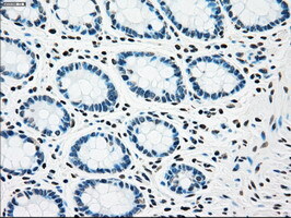
- Experimental details
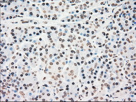
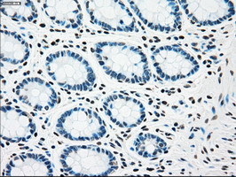
- IHC of paraffin-embedded colon tissue using anti-CHEK2 mouse monoclonal antibody. (Dilution 1:50).
- Submitted by
- LSBio (provider)
- Enhanced method
- Genetic validation
- Main image

- Experimental details
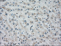
- IHC of paraffin-embedded liver tissue using anti-CHEK2 mouse monoclonal antibody. (Dilution 1:50).

- Submitted by
- LSBio (provider)
- Enhanced method
- Genetic validation
- Main image
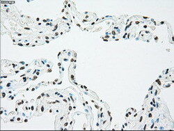
- Experimental details
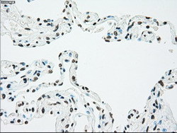
- IHC of paraffin-embedded lung tissue using anti-CHEK2 mouse monoclonal antibody. (Dilution 1:50).

- Submitted by
- LSBio (provider)
- Enhanced method
- Genetic validation
- Main image
- Experimental details
- IHC of paraffin-embedded pancreas tissue using anti-CHEK2 mouse monoclonal antibody. (Dilution 1:50).

- Submitted by
- LSBio (provider)
- Enhanced method
- Genetic validation
- Main image

- Experimental details
- IHC of paraffin-embedded colon tissue using anti-CHEK2 mouse monoclonal antibody. (Dilution 1:50).

- Submitted by
- LSBio (provider)
- Enhanced method
- Genetic validation
- Main image
- Experimental details
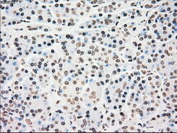
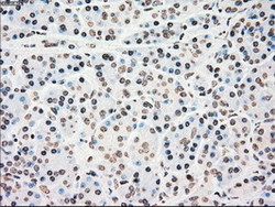
- IHC of paraffin-embedded Kidney tissue using anti-CHEK2 mouse monoclonal antibody. (Dilution 1:50).
- Submitted by
- LSBio (provider)
- Enhanced method
- Genetic validation
- Main image

- Experimental details
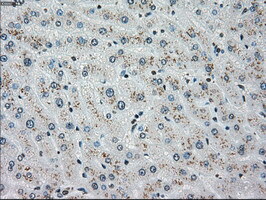
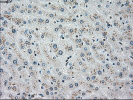
- IHC of paraffin-embedded liver tissue using anti-CHEK2 mouse monoclonal antibody. (Dilution 1:50).
- Submitted by
- LSBio (provider)
- Enhanced method
- Genetic validation
- Main image

- Experimental details
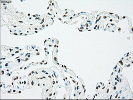
- IHC of paraffin-embedded lung tissue using anti-CHEK2 mouse monoclonal antibody. (Dilution 1:50).
- Submitted by
- LSBio (provider)
- Enhanced method
- Genetic validation
- Main image

- Experimental details
- IHC of paraffin-embedded pancreas tissue using anti-CHEK2 mouse monoclonal antibody. (Dilution 1:50).

- Submitted by
- LSBio (provider)
- Enhanced method
- Genetic validation
- Main image

- Experimental details
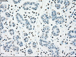
- IHC of paraffin-embedded breast tissue using anti-CHEK2 mouse monoclonal antibody. (Dilution 1:50).
- Submitted by
- LSBio (provider)
- Main image

- Experimental details
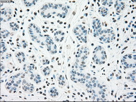
- IHC of paraffin-embedded breast tissue using anti-CHEK2 mouse monoclonal antibody. (Dilution 1:50).
- Submitted by
- LSBio (provider)
- Main image
- Experimental details
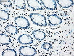
- IHC of paraffin-embedded colon tissue using anti-CHEK2 mouse monoclonal antibody. (Dilution 1:50).
- Submitted by
- LSBio (provider)
- Main image

- Experimental details
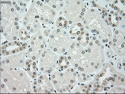
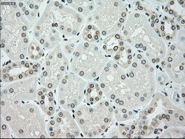
- IHC of paraffin-embedded Kidney tissue using anti-CHEK2 mouse monoclonal antibody. (Dilution 1:50).

- Submitted by
- LSBio (provider)
- Main image
- Experimental details
- IHC of paraffin-embedded liver tissue using anti-CHEK2 mouse monoclonal antibody. (Dilution 1:50).
- Submitted by
- LSBio (provider)
- Main image
- Experimental details
- IHC of paraffin-embedded lung tissue using anti-CHEK2 mouse monoclonal antibody. (Dilution 1:50).

- Submitted by
- LSBio (provider)
- Main image
- Experimental details
- IHC of paraffin-embedded pancreas tissue using anti-CHEK2 mouse monoclonal antibody. (Dilution 1:50).