Explore
 Validate
 Learn
LearnPA5-17818
antibody from Invitrogen Antibodies
Targeting: CHEK2
bA444G7, CDS1, CHK2, HuCds1, PP1425, RAD53
 Western blot
 Immunocytochemistry
Antibody data
- Antibody Data
- Antigen structure
- References [0]
- Comments [0]
- Validations
- Immunocytochemistry [6]
- Flow cytometry [2]
Submit
Validation data
Reference
Comment
Report error
- Product number
- PA5-17818 - Provider product page

- Provider
- Invitrogen Antibodies
- Product name
- Phospho-CHK2 (Thr68) Polyclonal Antibody
- Antibody type
- Polyclonal
- Antigen
- Synthetic peptide
- Description
- It is not recommended to aliquot this antibody. This antibody is not cross-reactive with Chk2 phosphorylated at other sites. This antibody was orginally validated as part of a Thermo Scientific Cellomics High Content Screening Kit. The antibody sold separately may have slightly different performance and may need to be further optimized for the best results.
- Reactivity
- Human
- Host
- Rabbit
- Isotype
- IgG
- Vial size
- 100 μL
- Concentration
- 99 μg/mL
- Storage
- -20°C
No comments: Submit comment
Supportive validation
- Submitted by
- Invitrogen Antibodies (provider)
- Main image
- Experimental details
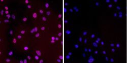
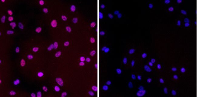
- (A) Chk2 phosphorylation: Chk2 is phosphorylated by ATM kinase in response to DNA damage and is important in DNA damage induced checkpoint signaling. A549 cells were treated with vehicle (DMSO) or 50 µM etoposide for 3 hrs. After staining cells with Cellomics Chk2 activation kit, images were acquired and analyzed. Z-factor for phospho-Chk2 is 0.49 ± 0.03 and %COV is 11.2 ± 0.4. EC50 of etoposide treatment for Chk2 phosphorylation is 1.8 µM.
- Submitted by
- Invitrogen Antibodies (provider)
- Main image
- Experimental details
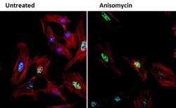
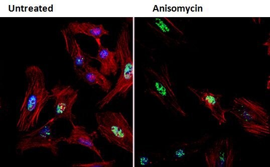
- Immunofluorescent analysis of Phospho-Chk2 pThr68 (green) in HeLa cells either left untreated (left panel) or treated with 25 µg/mL anisomycin for 30 minutes (right panel). Formalin fixed cells were permeabilized with 0.1% Triton X-100 in TBS for 10 minutes at room temperature and blocked with 1% Blocker BSA (Product # 37525) for 15 minutes at room temperature. Cells were probed with a Phospho-Chk2 pThr68 polyclonal antibody (Product # PA5-17818) at a dilution of 1:100 for at least 1 hour at room temperature, washed with PBS, and incubated with DyLight 488 goat anti-rabbit IgG secondary antibody (Product # 35552) at a dilution of 1:400 for 30 minutes at room temperature. F-Actin (red) was stained with DyLight 554 Phalloidin (Product # 21834) and nuclei (blue) were stained with Hoechst 33342 dye (Product # 62249). Images were taken on a Thermo Scientific ArrayScan or ToxInsight Instrument at 20X magnification.

- Submitted by
- Invitrogen Antibodies (provider)
- Main image

- Experimental details
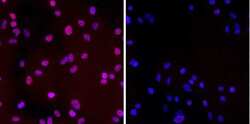
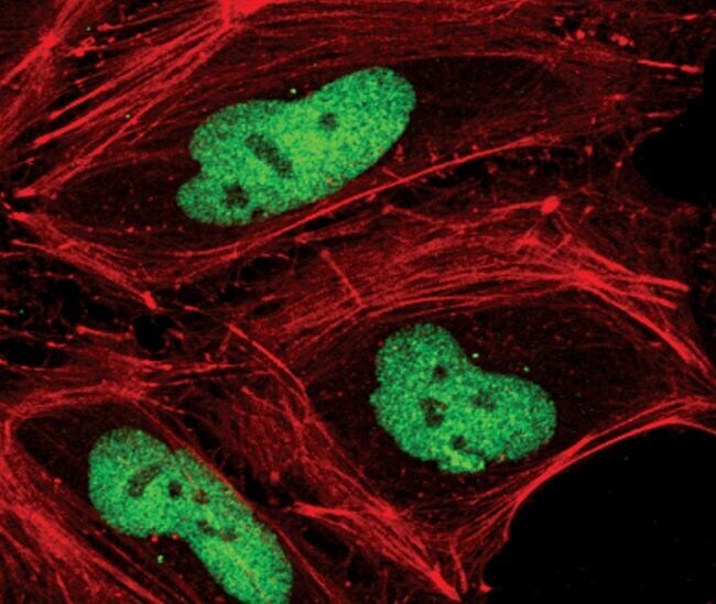
- Immunofluorescent analysis of phospho-Chk2 pThr68 (green) in UV-treated HeLa cells. Formaldehyde fixed cells were blocked in 5% normal goat serum + 0.3% Triton X-100 in PBS for 1 hour at room temperature. Cells were probed with a Phospho-Chk2 pThr68 polyclonal antibody (Product # PA5-17818) at a dilution of 1:100 overnight at 4C, followed by a fluorescently-conjugated anti-rabbit IgG secondary antibody. Actin (red) was stained using DyLight 554 Phalloidin (Product # 21834). Cells were visualized by confocal microscopy.
- Submitted by
- Invitrogen Antibodies (provider)
- Main image

- Experimental details
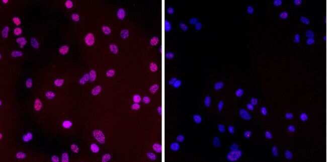
- Immunofluorescent analysis of phospho-Chk2 pThr68 (green) in UV-treated HeLa cells. Formaldehyde fixed cells were blocked in 5% normal goat serum + 0.3% Triton X-100 in PBS for 1 hour at room temperature. Cells were probed with a Phospho-Chk2 pThr68 polyclonal antibody (Product # PA5-17818) at a dilution of 1:100 overnight at 4C, followed by a fluorescently-conjugated anti-rabbit IgG secondary antibody. Actin (red) was stained using DyLight 554 Phalloidin (Product # 21834). Cells were visualized by confocal microscopy.

- Submitted by
- Invitrogen Antibodies (provider)
- Main image

- Experimental details
- Immunofluorescent analysis of Phospho-Chk2 pThr68 (green) in HeLa cells either left untreated (left panel) or treated with 25 µg/mL anisomycin for 30 minutes (right panel). Formalin fixed cells were permeabilized with 0.1% Triton X-100 in TBS for 10 minutes at room temperature and blocked with 1% Blocker BSA (Product # 37525) for 15 minutes at room temperature. Cells were probed with a Phospho-Chk2 pThr68 polyclonal antibody (Product # PA5-17818) at a dilution of 1:100 for at least 1 hour at room temperature, washed with PBS, and incubated with DyLight 488 goat anti-rabbit IgG secondary antibody (Product # 35552) at a dilution of 1:400 for 30 minutes at room temperature. F-Actin (red) was stained with DyLight 554 Phalloidin (Product # 21834) and nuclei (blue) were stained with Hoechst 33342 dye (Product # 62249). Images were taken on a Thermo Scientific ArrayScan or ToxInsight Instrument at 20X magnification.
- Submitted by
- Invitrogen Antibodies (provider)
- Main image
- Experimental details
- (A) Chk2 phosphorylation: Chk2 is phosphorylated by ATM kinase in response to DNA damage and is important in DNA damage induced checkpoint signaling. A549 cells were treated with vehicle (DMSO) or 50 µM etoposide for 3 hrs. After staining cells with Cellomics Chk2 activation kit, images were acquired and analyzed. Z-factor for phospho-Chk2 is 0.49 ± 0.03 and %COV is 11.2 ± 0.4. EC50 of etoposide treatment for Chk2 phosphorylation is 1.8 µM.
Supportive validation
- Submitted by
- Invitrogen Antibodies (provider)
- Main image

- Experimental details
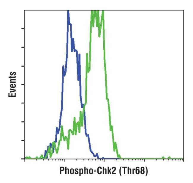
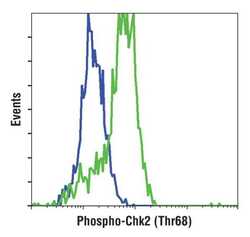
- Flow cytometric analysis of phospho-Chk2 pThr68 on untreated (blue histogram) or UV-treated (green histogram) HeLa cells. Cells were harvested, fixed with formaldehyde, permeabilized with methanol, and incubated with a Phospho-Chk2 pThr68 polyclonal antibody (Product # PA5-17818) at a 1:800 dilution for 1 hour at room temperature. Cells were stained with a fluorescently-conjugated anti-rabbit IgG secondary antibody and analyzed on a flow cytometer.

- Submitted by
- Invitrogen Antibodies (provider)
- Main image
- Experimental details
- Flow cytometric analysis of phospho-Chk2 pThr68 on untreated (blue histogram) or UV-treated (green histogram) HeLa cells. Cells were harvested, fixed with formaldehyde, permeabilized with methanol, and incubated with a Phospho-Chk2 pThr68 polyclonal antibody (Product # PA5-17818) at a 1:800 dilution for 1 hour at room temperature. Cells were stained with a fluorescently-conjugated anti-rabbit IgG secondary antibody and analyzed on a flow cytometer.