Explore
 Validate
 Learn
LearnNB100-56546
antibody from Novus Biologicals
Targeting: CHEK2
bA444G7, CDS1, CHK2, HuCds1, PP1425, RAD53
 Western blot
 Immunocytochemistry
Antibody data
- Antibody Data
- Antigen structure
- References [0]
- Comments [0]
- Validations
- Western blot [2]
- Immunohistochemistry [1]
- Flow cytometry [1]
Submit
Validation data
Reference
Comment
Report error
- Product number
- NB100-56546 - Provider product page

- Provider
- Novus Biologicals
- Proper citation
- Novus Cat#NB100-56546, RRID:AB_838021
- Product name
- Mouse Monoclonal Chk2 Antibody
- Antibody type
- Monoclonal
- Description
- Protein G purified.
- Reactivity
- Human, Mouse
- Host
- Mouse
- Isotype
- IgG
- Vial size
- 0.1 mg
- Concentration
- 0.5 mg/ml
- Storage
- Store at 4C short term. Aliquot and store at -20C long term. Avoid freeze-thaw cycles.
No comments: Submit comment
Supportive validation
- Submitted by
- Novus Biologicals (provider)
- Main image
- Experimental details
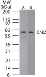
- Western Blot: Chk2 Antibody (73C175.1.1) [NB100-56546] - analysis of Chk2 in A) human HEK293 and B) mouse NIH3T3 lysate using Chk2 antibody at 2 ug/ml.

- Submitted by
- Novus Biologicals (provider)
- Main image
- Experimental details
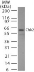
- Western Blot: Chk2 Antibody (73C175.1.1) [NB100-56546] - Western Blot: Chk2 Antibody (73C175.1.1) - Azide Free [NBP2-27403] - analysis of Chk2 in 293 cell lysate. A protein band of approximate molecular weight of 60-62 kDa is detected with Chk2 antibody at 2 ug/ml. Image using the Azide Free form of this antibody.
Supportive validation
- Submitted by
- Novus Biologicals (provider)
- Main image

- Experimental details
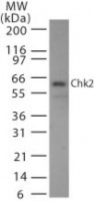
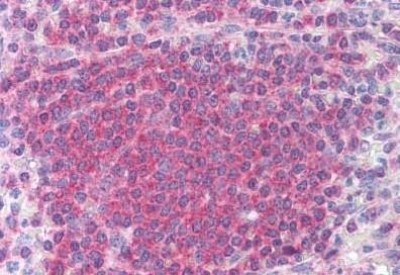
- Immunohistochemistry-Paraffin: Chk2 Antibody (73C175.1.1) [NB100-56546] - Staining of human spleen. Immunohistochemistry of formalin-fixed, paraffin-embedded tissue after heat-induced antigen retrieval. Antibody concentration 10 ug/ml.
Supportive validation
- Submitted by
- Novus Biologicals (provider)
- Main image
- Experimental details
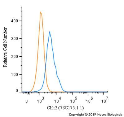
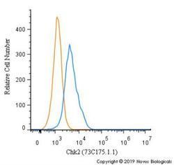
- Flow Cytometry: Chk2 Antibody (73C175.1.1) [NB100-56546] - An intracellular stain was performed on Hek293 cells with Chk2 [73C175.1.1] Antibody NB100-56546 (blue) and a matched isotype control (orange). Cells were fixed with 4% PFA and then permeabilized with 0.1% saponin. Cells were incubated in an antibody dilution of 2.5 ug/mL for 30 minutes at room temperature, followed by Mouse IgG (H+L) Cross-Adsorbed Secondary Antibody, Dylight 550.