Explore
 Validate
 Learn
Learn Western blot
Antibody data
- Antibody Data
- Antigen structure
- References [6]
- Comments [0]
- Validations
- Western blot [1]
- Immunocytochemistry [1]
Submit
Validation data
Reference
Comment
Report error
- Product number
- AF1626 - Provider product page

- Provider
- R&D Systems
- Product name
- Human Phospho-Chk2 (T68) Antibody
- Antibody type
- Polyclonal
- Description
- Antigen Affinity-purified. Detects human Chk2 when phosphorylated at T68.
- Reactivity
- Human
- Host
- Rabbit
- Conjugate
- Unconjugated
- Isotype
- IgG
- Vial size
- 100 ug
- Concentration
- LYOPH
- Storage
- Use a manual defrost freezer and avoid repeated freeze-thaw cycles. 12 months from date of receipt, -20 to -70 °C as supplied. 1 month, 2 to 8 °C under sterile conditions after reconstitution. 6 months, -20 to -70 °C under sterile conditions after reconstitution.
Submitted references Starvation-induced activation of ATM/Chk2/p53 signaling sensitizes cancer cells to cisplatin.
Multiple DNA damage signaling and repair pathways deregulated by simian virus 40 large T antigen.
RAD51C facilitates checkpoint signaling by promoting CHK2 phosphorylation.
Simian virus 40 large T antigen disrupts genome integrity and activates a DNA damage response via Bub1 binding.
Sonic Hedgehog signaling impairs ionizing radiation-induced checkpoint activation and induces genomic instability.
RNF8-dependent and RNF8-independent regulation of 53BP1 in response to DNA damage.
Shi Y, Felley-Bosco E, Marti TM, Orlowski K, Pruschy M, Stahel RA
BMC cancer 2012 Dec 4;12:571
BMC cancer 2012 Dec 4;12:571
Multiple DNA damage signaling and repair pathways deregulated by simian virus 40 large T antigen.
Boichuk S, Hu L, Hein J, Gjoerup OV
Journal of virology 2010 Aug;84(16):8007-20
Journal of virology 2010 Aug;84(16):8007-20
RAD51C facilitates checkpoint signaling by promoting CHK2 phosphorylation.
Badie S, Liao C, Thanasoula M, Barber P, Hill MA, Tarsounas M
The Journal of cell biology 2009 May 18;185(4):587-600
The Journal of cell biology 2009 May 18;185(4):587-600
Simian virus 40 large T antigen disrupts genome integrity and activates a DNA damage response via Bub1 binding.
Hein J, Boichuk S, Wu J, Cheng Y, Freire R, Jat PS, Roberts TM, Gjoerup OV
Journal of virology 2009 Jan;83(1):117-27
Journal of virology 2009 Jan;83(1):117-27
Sonic Hedgehog signaling impairs ionizing radiation-induced checkpoint activation and induces genomic instability.
Leonard JM, Ye H, Wetmore C, Karnitz LM
The Journal of cell biology 2008 Nov 3;183(3):385-91
The Journal of cell biology 2008 Nov 3;183(3):385-91
RNF8-dependent and RNF8-independent regulation of 53BP1 in response to DNA damage.
Sakasai R, Tibbetts R
The Journal of biological chemistry 2008 May 16;283(20):13549-55
The Journal of biological chemistry 2008 May 16;283(20):13549-55
No comments: Submit comment
Supportive validation
- Submitted by
- R&D Systems (provider)
- Main image

- Experimental details
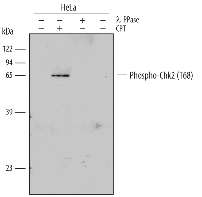
- Detection of Human Phospho-Chk2 (T68) by Western Blot. Western blot shows lysates of HeLa human cervical epithelial carcinoma cell line untreated (-) or treated (+) with 1 µM camptothecin (CPT) for 1 hour. PVDF membrane was probed with 0.5 µg/mL Rabbit Anti-Human Phospho-Chk2 (T68) Antigen Affinity-purified Polyclonal Antibody (Catalog # AF1626) followed by HRP-conjugated Anti-Rabbit IgG Secondary Antibody (Catalog # HAF008). A specific band for Phospho-Chk2 (T68) was detected at approximately 64 kDa (as indicated). The phospho-specificity of this antibody was supported by decreased labeling following treatment with 600 U lambda-phosphatase ( lambda-PPase) for 60 minutes. This experiment was conducted under reducing conditions and using Immunoblot Buffer Group 1.
Supportive validation
- Submitted by
- R&D Systems (provider)
- Main image

- Experimental details
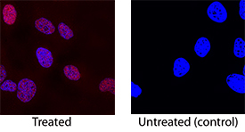
- Phospho-Chk2 (T68) in HepG2 Human Cell Line. Phopsho-Chk2 (T68) was detected in immersion fixed HepG2 human hepatocellular carcinoma cell line treated with 1 µM camptothecin using Rabbit Anti-Human Phospho-Chk2 (T68) Antigen Affinity-purified Polyclonal Antibody (Catalog # AF1626) at 1 µg/mL for 3 hours at room temperature. Cells were stained using the NorthernLights™ 557-conjugated Anti-Rabbit IgG Secondary Antibody (red; Catalog # NL004) and counterstained with DAPI (blue). Specific staining was localized to nuclei in treated cells. View our protocol for Fluorescent ICC Staining of Cells on Coverslips.