 Explore
 Validate
 Learn
Learn Western blot
 ELISA
ELISAAntibody data
- Antibody Data
- Antigen structure
- References [0]
- Comments [0]
- Validations
- Western blot [2]
Submit
Validation data
Reference
Comment
Report error
- Product number
- GTX48499 - Provider product page

- Provider
- GeneTex
- Proper citation
- GeneTex Cat#GTX48499, RRID:AB_11178208
- Product name
- GDF15 (H Variant) antibody [7C12.B3.F2]
- Antibody type
- Monoclonal
- Reactivity
- Human
- Host
- Mouse
No comments: Submit comment
Supportive validation
- Submitted by
- GeneTex (provider)
- Main image
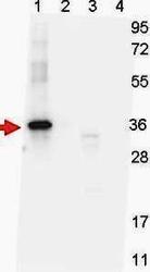
- Experimental details
- Western blot shows detection of recombinant NAG-1 protein present in Pichia pastoris whole cell lysates: lane 1 - yeast cell lysate expressing NAG-1 H variant with SUMO expression tag at 36 kDa; lane 2 - yeast cell lysate expressing NAG-1 D variant with SUMO expression tag at 36 kDa; lane 3 - yeast cell lysate expressing NAG-1 H variant; and lane 4 - yeast cell lysate expressing NAG-1 D variant. All lysates were run under reducing conditions. Primary antibody was used at a 1:1,000 dilution in TBS containing 1% BSA and 0.2% Tween, and reacted overnight at 4°C. For detection, a 1:40,000 dilution of peroxidase conjugated Gt-anti-Mouse IgG secondary antibody was used in Blocking Buffer for Fluorescent Western Blotting for 30 min at room temperature. Molecular weight estimation was made by comparison to prestained MW markers. Image was captured using the BioRad Versadoc 4000MP Imaging System.
- Validation comment
- WB

- Submitted by
- GeneTex (provider)
- Main image

- Experimental details
- Western blot shows detection of recombinant NAG-1 protein present in Pichia pastoris whole cell lysates: lane 1 - yeast cell lysate expressing NAG-1 H variant with SUMO expression tag at 36 kDa; lane 2 - yeast cell lysate expressing NAG-1 D variant with SUMO expression tag at 36 kDa; lane 3 - yeast cell lysate expressing NAG-1 H variant; and lane 4 - yeast cell lysate expressing NAG-1 D variant. All lysates were run under reducing conditions. Primary antibody was used at a 1:1,000 dilution in TBS containing 1% BSA and 0.2% Tween, and reacted overnight at 4¢XC. For detection, a 1:40,000 dilution of peroxidase conjugated Goat-anti-Mouse IgG secondary antibody for 30 min at room temperature. Molecular weight estimation was made by comparison to prestained MW markers.
