Explore
 Validate
 Learn
Learn Western blot
Antibody data
- Antibody Data
- Antigen structure
- References [2]
- Comments [0]
- Validations
- Western blot [1]
Submit
Validation data
Reference
Comment
Report error
- Product number
- MAB8161 - Provider product page

- Provider
- Abnova Corporation
- Proper citation
- Abnova Corporation Cat#MAB8161, RRID:AB_10676404
- Product name
- GDF15 monoclonal antibody, clone 23B3.D2.H5
- Antibody type
- Monoclonal
- Description
- Mouse monoclonal antibody raised against synthetic peptide of GDF15 .
- Isotype
- IgG
- Antibody clone number
- 23B3.D2.H5
- Storage
- Store at 4°C. For long term storage store at -20°C.Aliquot to avoid repeated freezing and thawing.
Submitted references Changes in gene expression contribute to cancer prevention by COX inhibitors.
H6D polymorphism in macrophage-inhibitory cytokine-1 gene associated with prostate cancer.
Baek SJ, Eling TE
Progress in lipid research 2006 Jan;45(1):1-16
Progress in lipid research 2006 Jan;45(1):1-16
H6D polymorphism in macrophage-inhibitory cytokine-1 gene associated with prostate cancer.
Lindmark F, Zheng SL, Wiklund F, Bensen J, Bälter KA, Chang B, Hedelin M, Clark J, Stattin P, Meyers DA, Adami HO, Isaacs W, Grönberg H, Xu J
Journal of the National Cancer Institute 2004 Aug 18;96(16):1248-54
Journal of the National Cancer Institute 2004 Aug 18;96(16):1248-54
No comments: Submit comment
Supportive validation
- Submitted by
- Abnova Corporation (provider)
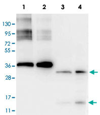
- Main image
- Experimental details
- Western blot using GDF15 monoclonal antibody, clone 23B3D2.H5 (Cat # MAB8161).The blot shows detection of recombinant GDF15 protein present in Pichia pastoris whole cell lysates.Lane 1, yeast cell lysate expressing GDF15 H variant with SUMO expression tag at 36 KDa.Lane 2, yeast cell lysate expressing GDF15 D variant with SUMO expression tag at 36 KDa.Lane 3, yeast cell lysate expressing GDF15 H variant.Lane 4, yeast cell lysate expressing GDF15 D variant.Recombinant GDF15 proteins without SUMO correspond to monomer (15 KDa) and dimer (30 KDa) bands as indicated by the arrowheads. All lysates were run under reducing conditions.Primary antibody was used at a 1 : 1,000 dilution in TBS containg 1% BSA and 0.2% Tween, and reacted overnight at 4°C. For detection, a 1 : 40,000 dilution of peroxidase conjugated Gt-a-Mouse IgG secondary antibody was used in Blocking Buffer for Fluorescent Western Blotting for 30 min at room temperature. Molecular weight estimation was made by comparison to prestained MW markers. Image was captured using the BioRad Versadoc™ 4000MP Imaging System.