 Explore
 Validate
 Learn
Learn Western blot
 Immunocytochemistry
Antibody data
- Antibody Data
- Antigen structure
- References [15]
- Comments [0]
- Validations
- Immunocytochemistry [1]
- Immunohistochemistry [1]
Submit
Validation data
Reference
Comment
Report error
- Product number
- HPA011191 - Provider product page

- Provider
- Atlas Antibodies
- Proper citation
- Atlas Antibodies Cat#HPA011191, RRID:AB_1078962
- Product name
- Anti-GDF15
- Antibody type
- Polyclonal
- Description
- Polyclonal Antibody against Human GDF15, Gene description: growth differentiation factor 15, Alternative Gene Names: MIC-1, MIC1, NAG-1, PDF, PLAB, PTGFB, Validated applications: WB, IHC, ICC, Uniprot ID: Q99988, Storage: Store at +4°C for short term storage. Long time storage is recommended at -20°C.
- Reactivity
- Human
- Host
- Rabbit
- Conjugate
- Unconjugated
- Isotype
- IgG
- Vial size
- 100 µl
- Concentration
- 0.1 mg/ml
- Storage
- Store at +4°C for short term storage. Long time storage is recommended at -20°C.
- Handling
- The antibody solution should be gently mixed before use.
Submitted references GDF15 propeptide promotes bone metastasis of castration-resistant prostate cancer by augmenting the bone microenvironment
GDF15 Promotes Cell Growth, Migration, and Invasion in Gastric Cancer by Inducing STAT3 Activation
Tumor-derived GDF-15 blocks LFA-1 dependent T cell recruitment and suppresses responses to anti-PD-1 treatment
Increased expression and accumulation of GDF15 in IPF extracellular matrix contribute to fibrosis
Expression of the Body-Weight Signaling Players: GDF15, GFRAL and RET and their clinical relevance in Gastric Cancer
Presence of growth/differentiation factor-15 cytokine in human follicular fluid, granulosa cells, and oocytes
Combined Secretomics and Transcriptomics Revealed Cancer-Derived GDF15 is Involved in Diffuse-Type Gastric Cancer Progression and Fibroblast Activation
GDF15 derived from both tumor-associated macrophages and esophageal squamous cell carcinomas contributes to tumor progression via Akt and Erk pathways
Reconstitution of TGFBR2-Mediated Signaling Causes Upregulation of GDF-15 in HCT116 Colorectal Cancer Cells
Association of Serum Level of Growth Differentiation Factor 15 with Liver Cirrhosis and Hepatocellular Carcinoma
The role of growth differentiation factor 15 in the pathogenesis of primary myelofibrosis.
GDF-15 is abundantly expressed in plexiform lesions in patients with pulmonary arterial hypertension and affects proliferation and apoptosis of pulmonary endothelial cells
Growth differentiation factor 15: a prognostic marker for recurrence in colorectal cancer.
Primary Ovarian Mucinous Carcinoma of Intestinal Type: Significance of Pattern of Invasion and Immunohistochemical Expression Profile in a Series of 31 Cases
GDF-15 Contributes to Proliferation and Immune Escape of Malignant Gliomas
Yamamichi G, Kato T, Arakawa N, Ino Y, Ujike T, Nakano K, Koh Y, Motoyama Y, Outani H, Myoba S, Ishizuya Y, Yamamoto Y, Hatano K, Kawashima A, Fukuhara S, Uemura H, Okada S, Morii E, Nonomura N, Uemura M
Biomarker Research 2024;12(1)
Biomarker Research 2024;12(1)
GDF15 Promotes Cell Growth, Migration, and Invasion in Gastric Cancer by Inducing STAT3 Activation
Joo M, Kim D, Lee M, Lee H, Kim J
International Journal of Molecular Sciences 2023;24(3):2925
International Journal of Molecular Sciences 2023;24(3):2925
Tumor-derived GDF-15 blocks LFA-1 dependent T cell recruitment and suppresses responses to anti-PD-1 treatment
Haake M, Haack B, Schäfer T, Harter P, Mattavelli G, Eiring P, Vashist N, Wedekink F, Genssler S, Fischer B, Dahlhoff J, Mokhtari F, Kuzkina A, Welters M, Benz T, Sorger L, Thiemann V, Almanzar G, Selle M, Thein K, Späth J, Gonzalez M, Reitinger C, Ipsen-Escobedo A, Wistuba-Hamprecht K, Eichler K, Filipski K, Zeiner P, Beschorner R, Goedemans R, Gogolla F, Hackl H, Rooswinkel R, Thiem A, Roche P, Joshi H, Pühringer D, Wöckel A, Diessner J, Rüdiger M, Leo E, Cheng P, Levesque M, Goebeler M, Sauer M, Nimmerjahn F, Schuberth-Wagner C, von Felten S, Mittelbronn M, Mehling M, Beilhack A, van der Burg S, Riedel A, Weide B, Dummer R, Wischhusen J
Nature Communications 2023;14(1)
Nature Communications 2023;14(1)
Increased expression and accumulation of GDF15 in IPF extracellular matrix contribute to fibrosis
Radwanska A, Cottage C, Piras A, Overed-Sayer C, Sihlbom C, Budida R, Wrench C, Connor J, Monkley S, Hazon P, Schluter H, Thomas M, Hogaboam C, Murray L
JCI Insight 2022;7(16)
JCI Insight 2022;7(16)
Expression of the Body-Weight Signaling Players: GDF15, GFRAL and RET and their clinical relevance in Gastric Cancer
Buchholz K, Antosik P, Grzanka D, Gagat M, Smolińska M, Grzanka A, Gzil A, Kasperska A, Klimaszewska-Wiśniewska A
Journal of Cancer 2021;12(15):4698-4709
Journal of Cancer 2021;12(15):4698-4709
Presence of growth/differentiation factor-15 cytokine in human follicular fluid, granulosa cells, and oocytes
Souček K, Malenovská A, Kahounová Z, Remšík J, Holubcová Z, Soukup T, Kurfürstová D, Bouchal J, Suchánková T, Slabáková E, Hampl A
Journal of Assisted Reproduction and Genetics 2018;35(8):1407-1417
Journal of Assisted Reproduction and Genetics 2018;35(8):1407-1417
Combined Secretomics and Transcriptomics Revealed Cancer-Derived GDF15 is Involved in Diffuse-Type Gastric Cancer Progression and Fibroblast Activation
Ishige T, Nishimura M, Satoh M, Fujimoto M, Fukuyo M, Semba T, Kado S, Tsuchida S, Sawai S, Matsushita K, Togawa A, Matsubara H, Kaneda A, Nomura F
Scientific Reports 2016;6(1)
Scientific Reports 2016;6(1)
GDF15 derived from both tumor-associated macrophages and esophageal squamous cell carcinomas contributes to tumor progression via Akt and Erk pathways
Urakawa N, Utsunomiya S, Nishio M, Shigeoka M, Takase N, Arai N, Kakeji Y, Koma Y, Yokozaki H
Laboratory Investigation 2015;95(5):491-503
Laboratory Investigation 2015;95(5):491-503
Reconstitution of TGFBR2-Mediated Signaling Causes Upregulation of GDF-15 in HCT116 Colorectal Cancer Cells
Sun L, Lee J, Fricke F, Warnken U, Schnölzer M, Kopitz J, Gebert J
PLOS ONE 2015;10(6):e0131506
PLOS ONE 2015;10(6):e0131506
Association of Serum Level of Growth Differentiation Factor 15 with Liver Cirrhosis and Hepatocellular Carcinoma
Zhang L, Liu X, Chi X, Gong Q, Gao L, Niu Y, Chi X, Cheng M, Si Y, Wang M, Zhong J, Niu J, Yang W
PLOS ONE 2015;10(5):e0127518
PLOS ONE 2015;10(5):e0127518
The role of growth differentiation factor 15 in the pathogenesis of primary myelofibrosis.
Uchiyama T, Kawabata H, Miura Y, Yoshioka S, Iwasa M, Yao H, Sakamoto S, Fujimoto M, Haga H, Kadowaki N, Maekawa T, Takaori-Kondo A
Cancer medicine 2015 Oct;4(10):1558-72
Cancer medicine 2015 Oct;4(10):1558-72
GDF-15 is abundantly expressed in plexiform lesions in patients with pulmonary arterial hypertension and affects proliferation and apoptosis of pulmonary endothelial cells
Nickel N, Jonigk D, Kempf T, Bockmeyer C, Maegel L, Rische J, Laenger F, Lehmann U, Sauer C, Greer M, Welte T, Hoeper M, Golpon H
Respiratory Research 2011;12(1)
Respiratory Research 2011;12(1)
Growth differentiation factor 15: a prognostic marker for recurrence in colorectal cancer.
Wallin U, Glimelius B, Jirström K, Darmanis S, Nong RY, Pontén F, Johansson C, Påhlman L, Birgisson H
British journal of cancer 2011 May 10;104(10):1619-27
British journal of cancer 2011 May 10;104(10):1619-27
Primary Ovarian Mucinous Carcinoma of Intestinal Type: Significance of Pattern of Invasion and Immunohistochemical Expression Profile in a Series of 31 Cases
Tabrizi A, Kalloger S, Köbel M, Cipollone J, Roskelley C, Mehl E, Gilks C
International Journal of Gynecological Pathology 2010;29(2):99-107
International Journal of Gynecological Pathology 2010;29(2):99-107
GDF-15 Contributes to Proliferation and Immune Escape of Malignant Gliomas
Roth P, Junker M, Tritschler I, Mittelbronn M, Dombrowski Y, Breit S, Tabatabai G, Wick W, Weller M, Wischhusen J
Clinical Cancer Research 2010;16(15):3851-3859
Clinical Cancer Research 2010;16(15):3851-3859
No comments: Submit comment
Supportive validation
- Submitted by
- Atlas Antibodies (provider)
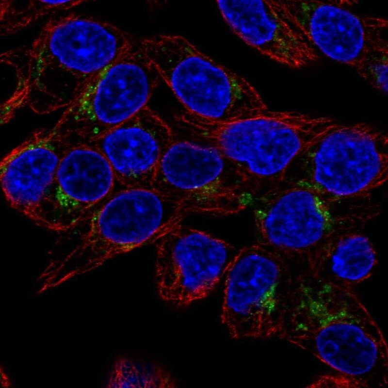
- Main image
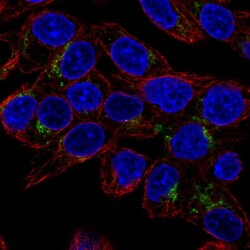
- Experimental details
- Immunofluorescent staining of human cell line Hep G2 shows localization to the Golgi apparatus.
- Sample type
- Human
Supportive validation
- Submitted by
- Atlas Antibodies (provider)
- Enhanced method
- Orthogonal validation
- Main image
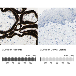
- Experimental details
- Immunohistochemistry analysis in human placenta and cervix, uterine tissues using HPA011191 antibody. Corresponding GDF15 RNA-seq data are presented for the same tissues.
- Sample type
- Human
- Protocol
- Protocol
