Explore
 Validate
 Learn
Learn Western blot
Antibody data
- Antibody Data
- Antigen structure
- References [3]
- Comments [0]
- Validations
- Western blot [6]
- Immunocytochemistry [4]
- Immunoprecipitation [1]
- Immunohistochemistry [4]
- Other assay [5]
Submit
Validation data
Reference
Comment
Report error
- Product number
- PA5-34772 - Provider product page

- Provider
- Invitrogen Antibodies
- Product name
- HSPA1A Polyclonal Antibody
- Antibody type
- Polyclonal
- Antigen
- Recombinant protein fragment
- Description
- Recommended positive controls: 293T, NIH-3T3, Mouse brain, PC-12, Rat brain, Jurkat, Raji, 293T, A431, HeLa, HepG2, H1299, HCT116, MCF-7, NT2D1, PC-3, U87-MG, Mesenchymal stem cells. Predicted reactivity: Mouse (97%), Rat (98%), Xenopus laevis (85%), Sheep (99%), Bovine (99%). Store product as a concentrated solution. Centrifuge briefly prior to opening the vial.
- Reactivity
- Human, Mouse, Rat
- Host
- Rabbit
- Isotype
- IgG
- Vial size
- 100 µL
- Concentration
- 0.62 mg/mL
- Storage
- Store at 4°C short term. For long term storage, store at -20°C, avoiding freeze/thaw cycles.
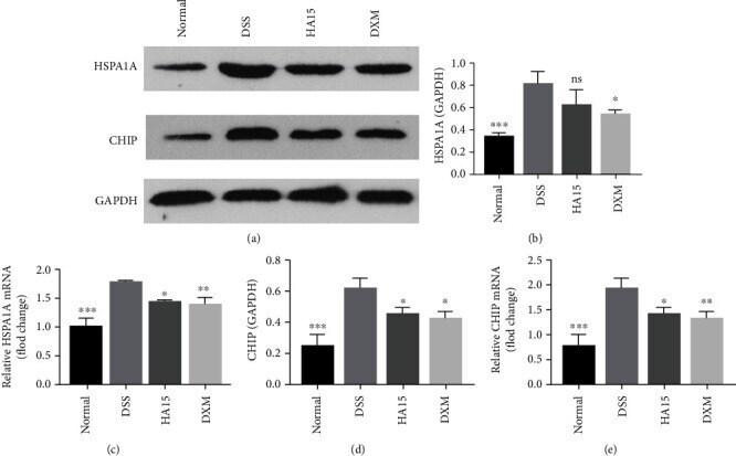
Submitted references HSPA5 Inhibitor Meliorate DSS-Induced Colitis through HSPA1A/CHIP.
Heat Shock Protein A6 Is Especially Involved in Enterovirus 71 Infection.
Age-associated changes in microglia and astrocytes ameliorate blood-brain barrier dysfunction.
Gao F, Fan H, Xue L, Shou Z, Zhu F, Yu T, Chu S, Wei C, Liu C, Zuo D, Zuo D
Disease markers 2022;2022:7115181
Disease markers 2022;2022:7115181
Heat Shock Protein A6 Is Especially Involved in Enterovirus 71 Infection.
Jia J, Liu G, Zhong J, Yan R, Song X, Zheng K, Ren Z, He Z, Zhu Q
Frontiers in microbiology 2022;13:865644
Frontiers in microbiology 2022;13:865644
Age-associated changes in microglia and astrocytes ameliorate blood-brain barrier dysfunction.
Pan J, Ma N, Zhong J, Yu B, Wan J, Zhang W
Molecular therapy. Nucleic acids 2021 Dec 3;26:970-986
Molecular therapy. Nucleic acids 2021 Dec 3;26:970-986
No comments: Submit comment
Supportive validation
- Submitted by
- Invitrogen Antibodies (provider)
- Main image
- Experimental details
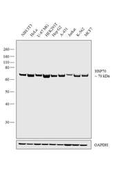
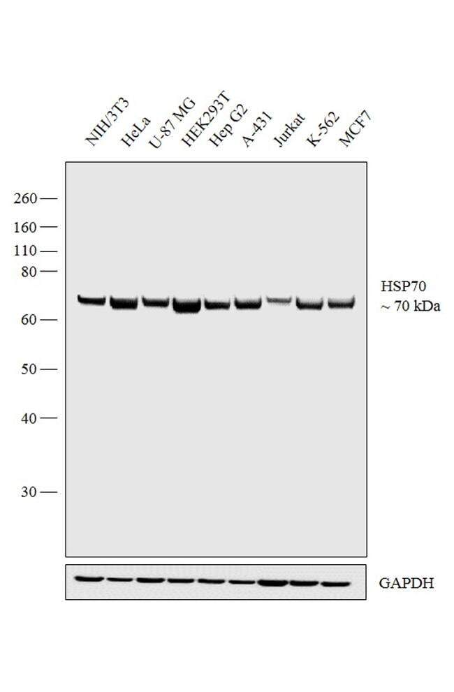
- Western blot analysis was performed on whole cell extracts (30 µg lysate) of NIH/3T3 (Lane 1), HeLa (Lane 2), U-87 MG (Lane 3), HEK293T (Lane 4), Hep G2 (Lane 5), A-431 (Lane 6), Jurkat (Lane 7), K-562 (Lane 8) and MCF7 (Lane 9). The blot was probed with Anti-HSP70 Polyclonal Antibody (Product # PA5-34772, 1 µg/mL) and detected by chemiluminescence using Goat anti-Rabbit IgG (H+L) Superclonal™ Secondary Antibody, HRP conjugate (Product # A27036, 0.25 µg/mL, 1:4000 dilution). A 70 kDa band corresponding to HSP70 was observed across the cell lines tested.
- Submitted by
- Invitrogen Antibodies (provider)
- Main image
- Experimental details
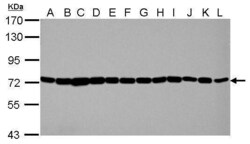
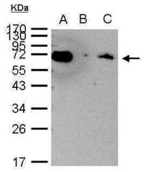
- Western Blot using HSPA1A Polyclonal Antibody (Product # PA5-34772). Sample (30 µg of whole cell lysate). Lane A: Jurkat. Lane B: Raji. Lane C: 293T. Lane D: A431. Lane E: HeLa. Lane F: HepG2. Lane G: H1299. Lane H: HCT116. I: MCF-7. J: NT2D1. K: PC-3. L: U87-MG. 7.5% SDS PAGE. HSPA1A Polyclonal Antibody (Product # PA5-34772) diluted at 1:10,000. The HRP-conjugated anti-rabbit IgG antibody was used to detect the primary antibody.

- Submitted by
- Invitrogen Antibodies (provider)
- Main image
- Experimental details
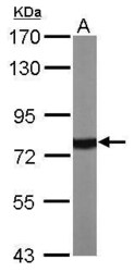
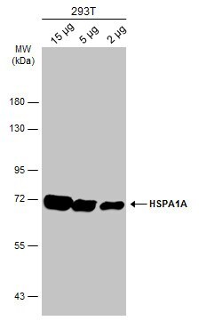
- Western Blot using HSPA1A Polyclonal Antibody (Product # PA5-34772). Sample (50 µg of whole cell lysate). Lane A: Mouse brain. 7.5% SDS PAGE. HSPA1A Polyclonal Antibody (Product # PA5-34772) diluted at 1:10,000. The HRP-conjugated anti-rabbit IgG antibody was used to detect the primary antibody.

- Submitted by
- Invitrogen Antibodies (provider)
- Main image
- Experimental details
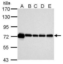
- Western Blot using HSPA1A Polyclonal Antibody (Product # PA5-34772). Sample (30 µg of whole cell lysate). Lane A: 293T. Lane B: NIH-3T3. Lane C: Mouse brain. Lane D: PC-12. Lane E: Rat brain. 7.5% SDS PAGE. HSPA1A Polyclonal Antibody (Product # PA5-34772) diluted at 1:10,000. The HRP-conjugated anti-rabbit IgG antibody was used to detect the primary antibody.
- Submitted by
- Invitrogen Antibodies (provider)
- Main image
- Experimental details
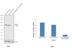
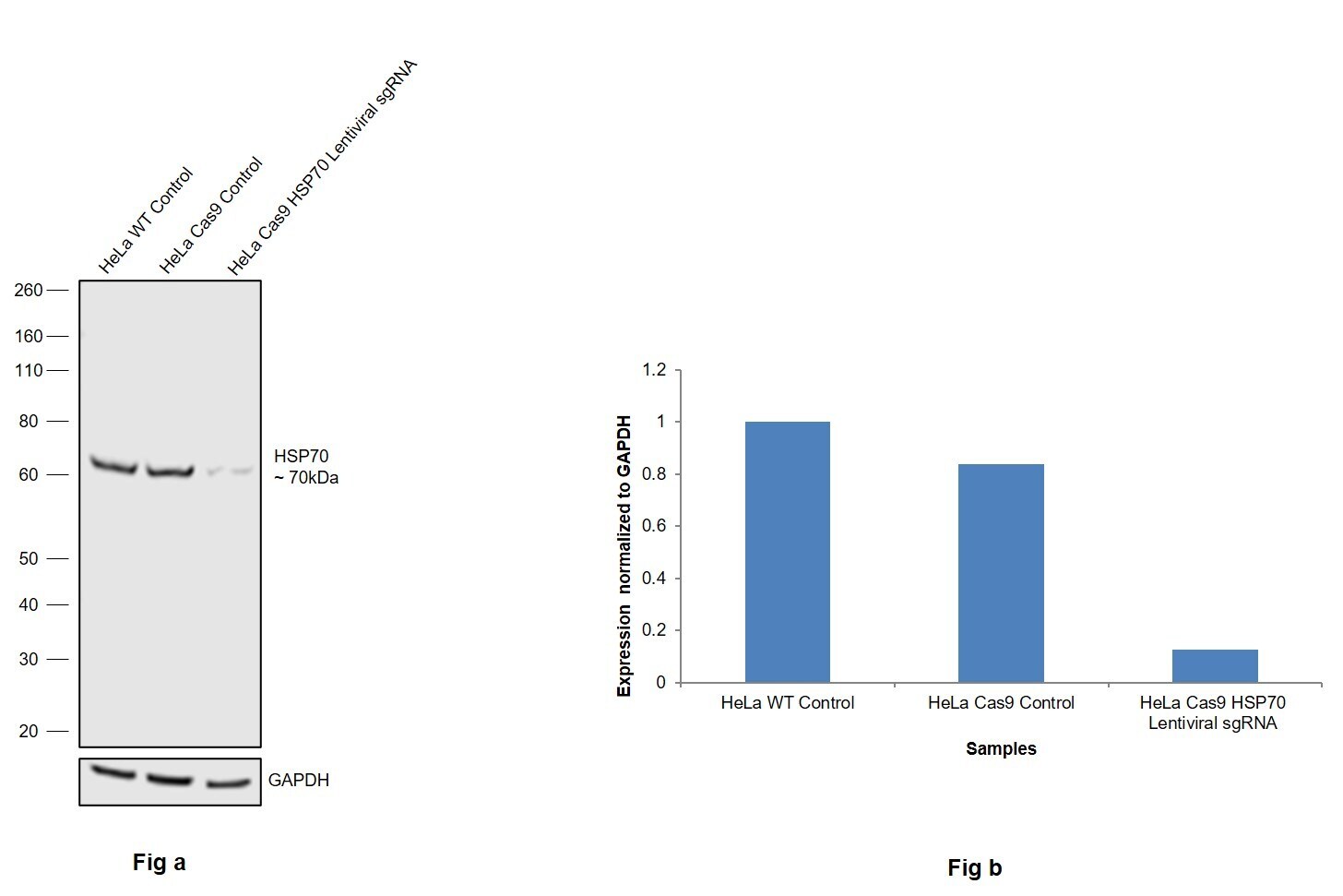
- CRISPR-Cas9 mediated genome editing ofHSP70 (as confirmed by next generation sequencing) was achieved by using LentiArray™ Lentiviral sgRNA (Product # A32042, AssayID CRISPR697585_LV) and LentiArray Cas9 Lentivirus (Product # A32064). Fig (a) Western blot analysis of HSP70 was performed by loading 30 µg of HeLa Wild Type (Lane 1), HeLa Cas9 (Lane 2) and HeLa Cas9 cells transduced with HSP70 Lentiviral sgRNA (Lane 3) whole cell extracts. The samples were electrophoresed using NuPAGE™ Novex™ 4-12% Bis-Tris Protein Gel (Product # NP0322BOX). Resolved proteins were then transferred onto a nitrocellulose membrane (Product # IB23001) by iBlot® 2 Dry Blotting System (Product # IB21001). The blot was probed with Anti-HSPA1A Polyclonal Antibody (Product # PA5-34772) using 1 µg/mL dilution and Goat anti-Rabbit IgG (Heavy Chain) Superclonal™ Recombinant Secondary Antibody, HRP (Product # A27036 1:5,000 dilution).Chemiluminescent detection was performed using Novex® ECL Chemiluminescent Substrate Reagent Kit (Product # WP20005). A reduced signal in sgRNA transduced cells using the LentiArray™ CRISPR product line confirms that antibody is specific toHSP70 (Fig (b)).
- Submitted by
- Invitrogen Antibodies (provider)
- Main image
- Experimental details
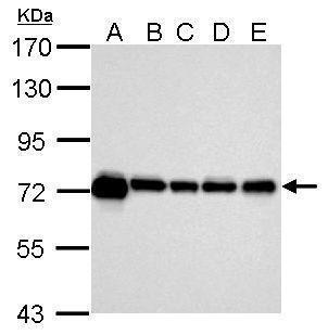
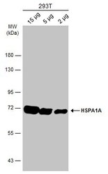
- Western Blot analysis of HSPA1 was performed by separating various whole cell extracts by 7.5% SDS-PAGE. Proteins were transferred to a membrane and probed with a HSPA1 Polyclonal Antibody (Product # PA5-34772) at a dilution of 1:10000 and a HRP-conjugated anti-rabbit IgG secondary antibody.
Supportive validation
- Submitted by
- Invitrogen Antibodies (provider)
- Main image
- Experimental details
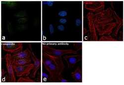
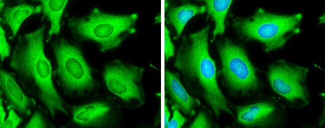
- Immunofluorescence analysis of HSP70 was performed using 70% confluent log phase HeLa cells. The cells were fixed with 4% paraformaldehyde for 10 minutes, permeabilized with 0.1% Triton™ X-100 for 10 minutes, and blocked with 1% BSA for 1 hour at room temperature. The cells were labeled with HSP70 Polyclonal Antibody (Product # PA5-34772) at 5 µg/mL in 0.1% BSA and incubated overnight at 4 degree and then labeled with Goat anti-Rabbit IgG (H+L) Superclonal™ Secondary Antibody, Alexa Fluor® 488 conjugate (Product # A27034) at a dilution of 1:2000 for 45 minutes at room temperature (Panel a: green). Nuclei (Panel b: blue) were stained with SlowFade® Gold Antifade Mountant with DAPI (Product # S36938). F-actin (Panel c: red) was stained with Rhodamine Phalloidin (Product # R415, 1:300). Panel d represents the merged image showing nuclear and cytoplasmic localization. Panel e represents control cells with no primary antibody to assess background. The images were captured at 60X magnification.

- Submitted by
- Invitrogen Antibodies (provider)
- Main image
- Experimental details
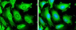
- Immunocytochemistry-Immunofluorescence analysis of HSPA1 was performed in HeLa cells fixed in ice-cold MeOH for 5 min. Green: HSPA1 Polyclonal Antibody (Product # PA5-34772) diluted at 1:500. Blue: Hoechst 33342 staining.

- Submitted by
- Invitrogen Antibodies (provider)
- Main image

- Experimental details
- Immunocytochemistry-Immunofluorescence analysis of HSPA1 was performed in HeLa cells fixed in ice-cold MeOH for 5 min. Green: HSPA1 Polyclonal Antibody (Product # PA5-34772) diluted at 1:500. Blue: Hoechst 33342 staining.
- Submitted by
- Invitrogen Antibodies (provider)
- Main image

- Experimental details
- Immunofluorescence analysis of HSP70 was performed using 70% confluent log phase HeLa cells. The cells were fixed with 4% paraformaldehyde for 10 minutes, permeabilized with 0.1% Triton™ X-100 for 10 minutes, and blocked with 1% BSA for 1 hour at room temperature. The cells were labeled with HSP70 Polyclonal Antibody (Product # PA5-34772) at 5 µg/mL in 0.1% BSA and incubated overnight at 4 degree and then labeled with Goat anti-Rabbit IgG (Heavy Chain) Superclonal™ Secondary Antibody, Alexa Fluor® 488 conjugate (Product # A27034) at a dilution of 1:2000 for 45 minutes at room temperature (Panel a: green). Nuclei (Panel b: blue) were stained with SlowFade® Gold Antifade Mountant with DAPI (Product # S36938). F-actin (Panel c: red) was stained with Rhodamine Phalloidin (Product # R415, 1:300). Panel d represents the merged image showing nuclear and cytoplasmic localization. Panel e represents control cells with no primary antibody to assess background. The images were captured at 60X magnification.

Supportive validation
- Submitted by
- Invitrogen Antibodies (provider)
- Main image

- Experimental details
- HSPA1A Polyclonal Antibody immunoprecipitates Hsp70 protein in IP experiments. IP Sample: 1,000 µg HeLa whole cell lysate/extract A. 40 µg HeLa whole cell lysate/extract B. Control with 2.5 µg of preimmune rabbit IgG C. Immunoprecipitation of Hsp70 protein by 2.5 µg of HSPA1A Polyclonal Antibody (Product # PA5-34772) 12% SDS-PAGE The immunoprecipitated Hsp70 protein was detected by HSPA1A Polyclonal Antibody (Product # PA5-34772) diluted at 1:1,000.

Supportive validation
- Submitted by
- Invitrogen Antibodies (provider)
- Main image
- Experimental details
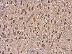
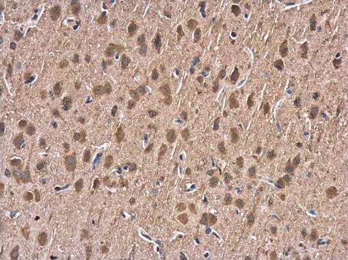
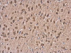
- Immunohistochemistry (Paraffin) analysis of HSPA1 was performed in paraffin-embedded rat brain tissue using HSPA1 Polyclonal Antibody (Product # PA5-34772) at a dilution of 1:500.
- Submitted by
- Invitrogen Antibodies (provider)
- Main image
- Experimental details
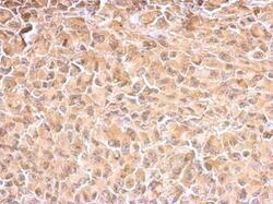
- HSPA1A Polyclonal Antibody detects Hsp70 protein at cytosol on H1299 xenograft by immunohistochemical analysis. Sample: Paraffin-embedded H1299 xenograft. HSPA1A Polyclonal Antibody (Product # PA5-34772) dilution: 1:500. Antigen Retrieval: EDTA based buffer, pH 8.0, 15 min.

- Submitted by
- Invitrogen Antibodies (provider)
- Main image

- Experimental details
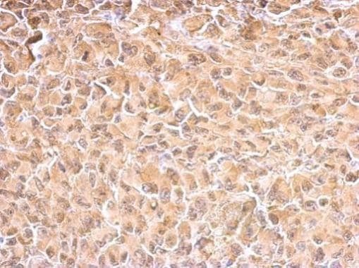
- HSPA1A Polyclonal Antibody detects Hsp70 protein at cytosol on H1299 xenograft by immunohistochemical analysis. Sample: Paraffin-embedded H1299 xenograft. HSPA1A Polyclonal Antibody (Product # PA5-34772) dilution: 1:500. Antigen Retrieval: EDTA based buffer, pH 8.0, 15 min.
- Submitted by
- Invitrogen Antibodies (provider)
- Main image
- Experimental details
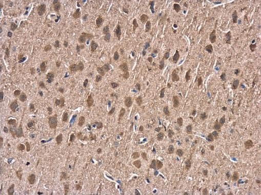
- Immunohistochemistry (Paraffin) analysis of HSPA1 was performed in paraffin-embedded rat brain tissue using HSPA1 Polyclonal Antibody (Product # PA5-34772) at a dilution of 1:500.
Supportive validation
- Submitted by
- Invitrogen Antibodies (provider)
- Main image
- Experimental details
- HSPA1A Polyclonal Antibody immunoprecipitates Hsp70 protein in IP experiments. IP Sample: 1,000 µg HeLa whole cell lysate/extract A. 40 µg HeLa whole cell lysate/extract B. Control with 2.5 µg of preimmune rabbit IgG C. Immunoprecipitation of Hsp70 protein by 2.5 µg of HSPA1A Polyclonal Antibody (Product # PA5-34772) 12% SDS-PAGE The immunoprecipitated Hsp70 protein was detected by HSPA1A Polyclonal Antibody (Product # PA5-34772) diluted at 1:1,000.

- Submitted by
- Invitrogen Antibodies (provider)
- Main image
- Experimental details
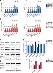
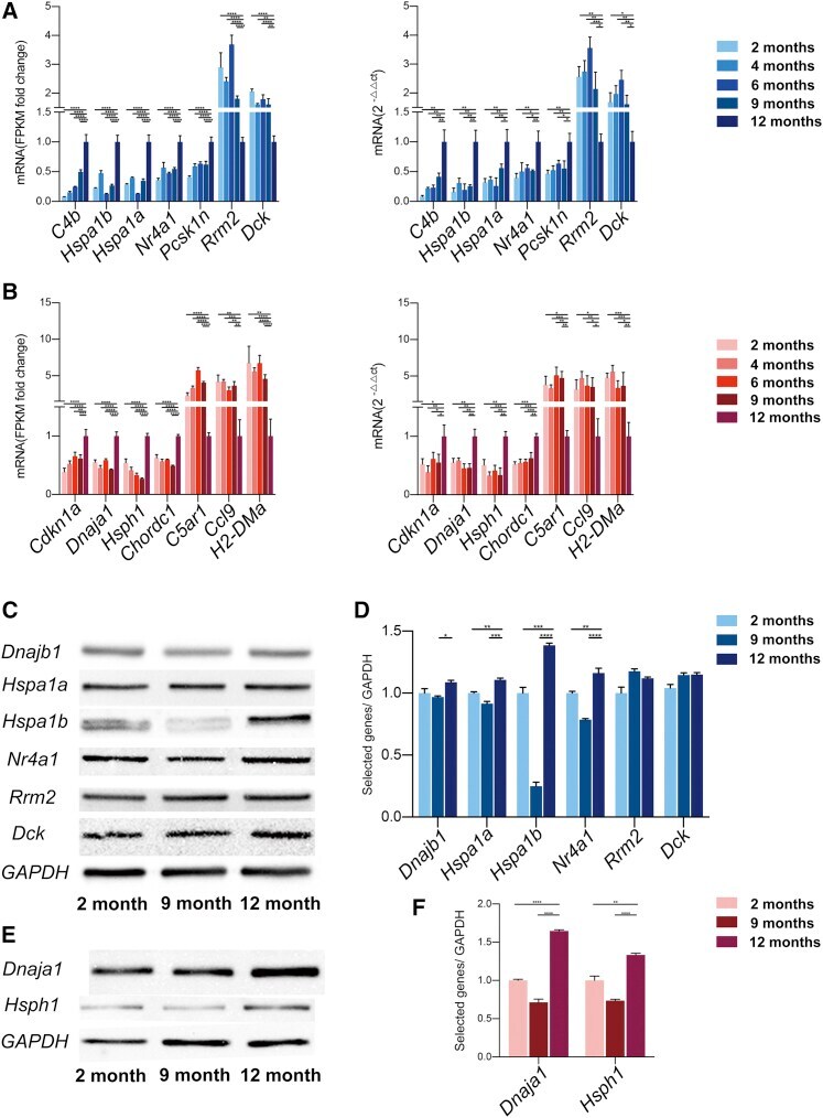
- Figure 8 Validation of transcriptome data by using qPCR and western blot (WB) (A and B) Expression analyses performed on selected genes yielded results superimposable with results obtained from RNA-seq analyses of (A) astrocytes and (B) microglia. Left: comparisons of DESeq2-analysis values between 12-month-olkd samples and 2-, 4-, 6-, and 9-month-old samples. Right: unpaired t tests for comparing two samples. (C) WB analysis of selected genes in astrocytic lysates. Loading was quantified by blotting with an anti-GAPDH antibody in cell lysates. (D) Quantification of the blot in (C) (unpaired t tests for comparing two samples). (E) WB analysis of selected genes in microglial lysates. Loading was quantified by blotting with an anti-GAPDH antibody in cell lysates. (F) Quantification of the blot in (E) (unpaired t tests for comparing two samples). Columns represent means +- SEM; ****p < 0.0001, ***p < 0.001, **p < 0.01, *p < 0.05.
- Submitted by
- Invitrogen Antibodies (provider)
- Main image

- Experimental details
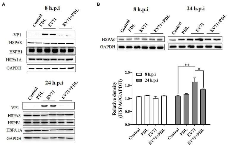
- Figure 2 EV71 infection specially upregulates HSPA6 protein expression. (A) Western blotting analysis of HSPA1A, HSPA8, HSPB1, and viral VP1 expression in RD cells infected with EV71 for 8 or 24 h. (B) Western blotting analysis of HSPA6 expression in RD cells infected with EV71 for 8 or 24 h. The relative expression of HSPA6 was quantitatively analyzed based on the bands' intensities from the Western blotting analysis. PDL (10 muM) here was used as anti-EV71 control ( * p < 0.05, ** p < 0.01).
- Submitted by
- Invitrogen Antibodies (provider)
- Main image
- Experimental details
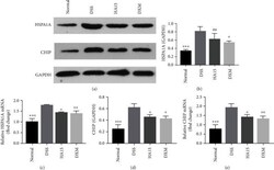
- Expressions of HSPA1A and CHIP in colonic tissue. (a) The colonic protein levels of HSPA1A and CHIP measured by Western blotting. (b) Representative protein levels of HSPA1A/GAPDH. (c) Levels of HSPA1A mRNA in colonic tissue. (d) Representative protein levels of CHIP/GAPDH. (e) Levels of CHIP mRNA in colonic tissue. Data are shown as means +- SD ( n = 3). ** P < 0.01, *** P < 0.001, vs. DSS group.