Explore
 Validate
 Learn
Learn Western blot
 Immunocytochemistry
Antibody data
- Antibody Data
- Antigen structure
- References [2]
- Comments [0]
- Validations
- Western blot [1]
Submit
Validation data
Reference
Comment
Report error
- Product number
- M00949-2 - Provider product page

- Provider
- Boster Biological Technology
- Product name
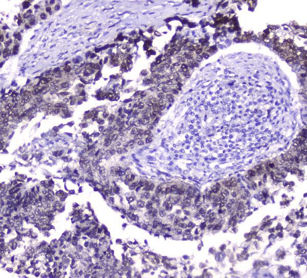
- Anti-Hsp70 HSPA1A Antibody Picoband™ (monoclonal, 3H5)
- Antibody type
- Monoclonal
- Description
- Mouse IgG monoclonal antibody for Hsp70 detection. Tested with WB, IHC-P, ICC/IF, FCM in Human;Mouse;Rat.
- Reactivity
- Human, Mouse, Rat
- Host
- Mouse
- Isotype
- IgG
- Antibody clone number
- 3H5
- Vial size
- 100μg/vial
- Concentration
- 0.5-1mg/ml, actual concentration vary by lot. Use suggested dilution ratio to decide dilution procedure.
- Storage
- At -20℃ for one year. After reconstitution, at 4℃ for one month. It can also be aliquoted and stored frozen at -20℃ for a longer time. Avoid repeated freezing and thawing.
- Handling
- Add 0.2ml of distilled water will yield a concentration of 500ug/ml.
Submitted references Study on the immune response to recombinant Hsp70 protein from Megalobrama amblycephala.
Down-modulation of heat shock protein 70 and up-modulation of Caspase-3 during schisandrin B-induced apoptosis in human hepatoma SMMC-7721 cells.
Chen N, Wan XL, Huang CX, Wang WM, Liu H, Wang HL
Immunobiology 2014 Nov;219(11):850-8
Immunobiology 2014 Nov;219(11):850-8
Down-modulation of heat shock protein 70 and up-modulation of Caspase-3 during schisandrin B-induced apoptosis in human hepatoma SMMC-7721 cells.
Wu YF, Cao MF, Gao YP, Chen F, Wang T, Zumbika EP, Qian KX
World journal of gastroenterology 2004 Oct 15;10(20):2944-8
World journal of gastroenterology 2004 Oct 15;10(20):2944-8
No comments: Submit comment
Supportive validation
- Submitted by
- Boster Biological Technology (provider)
- Main image
- Experimental details
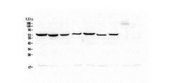
- Western blot analysis of Hsp70 using anti-Hsp70 antibody (M00949-2). Electrophoresis was performed on a 5-20% SDS-PAGE gel at 70V (Stacking gel) / 90V (Resolving gel) for 2-3 hours. The sample well of each lane was loaded with 50ug of sample under reducing conditions. Lane 1: human Hela whole cell lysate,Lane 2: human COLO-320 whole cell lysate,Lane 3: human SW620 whole cell lysate,Lane 4: human A431 whole cell lysate,Lane 5: human A549 whole cell lysate,Lane 6: human HepG2 whole cell lysate,Lane 7: human PANC-1 whole cell lysate. After Electrophoresis, proteins were transferred to a Nitrocellulose membrane at 150mA for 50-90 minutes. Blocked the membrane with 5% Non-fat Milk/ TBS for 1.5 hour at RT. The membrane was incubated with mouse anti-Hsp70 antigen affinity purified monoclonal antibody (Catalog # M00949-2) at 0.5 μg/mL overnight at 4°C, then washed with TBS-0.1%Tween 3 times with 5 minutes each and probed with a goat anti-mouse IgG-HRP secondary antibody at a dilution of 1:10000 for 1.5 hour at RT. The signal is developed using an Enhanced Chemiluminescent detection (ECL) kit (Catalog # EK1001) with Tanon 5200 system.
- Additional image