Explore
 Validate
 Learn
Learn Western blot
Antibody data
- Antibody Data
- Antigen structure
- References [1]
- Comments [0]
- Validations
- Western blot [4]
- Immunocytochemistry [1]
- Immunohistochemistry [1]
Submit
Validation data
Reference
Comment
Report error
- Product number
- GTX112584 - Provider product page

- Provider
- GeneTex
- Proper citation
- GeneTex Cat#GTX112584, RRID:AB_2037165
- Product name
- HHIP antibody [N3C2], Internal
- Antibody type
- Polyclonal
- Reactivity
- Human, Mouse, Chicken/Avian
- Host
- Rabbit
Submitted references Molecular analysis of the TGF-beta controlled gene expression program in chicken embryo dermal myofibroblasts.
Kosla J, Dvorak M, Cermak V
Gene 2013 Jan 15;513(1):90-100
Gene 2013 Jan 15;513(1):90-100
No comments: Submit comment
Supportive validation
- Submitted by
- GeneTex (provider)
- Main image
- Experimental details
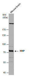
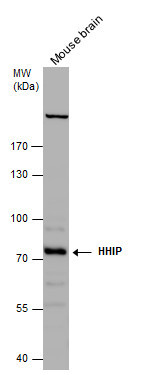
- Sample (50 ug of whole cell lysate) A: Mouse brain 7.5% SDS PAGE GTX112584 diluted at 1:1000
- Validation comment
- WB

- Submitted by
- GeneTex (provider)
- Main image

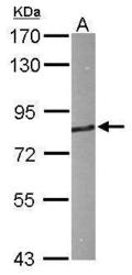
- Experimental details
- Sample (30 ug of whole cell lysate) A: A431 (GTX27909) 7.5% SDS PAGE GTX112584 diluted at 1:1000
- Validation comment
- WB
- Submitted by
- GeneTex (provider)
- Main image
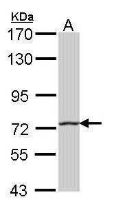
- Experimental details
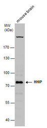
- HHIP antibody [N3C2], Internal detects HHIP protein by western blot analysis. Mouse tissue extracts (50 ?g) was separated by 7.5% SDS-PAGE, and the membrane was blotted with HHIP antibody [N3C2], Internal (GTX112584) diluted at 1:1000. The HRP-conjugated anti-rabbit IgG antibody (GTX213110-01) was used to detect the primary antibody.
- Submitted by
- GeneTex (provider)
- Main image
- Experimental details
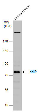
- HHIP antibody detects HHIP protein by Western blot analysis. Mouse tissue extracts (50 £gg) was separated by 7.5 % SDS-PAGE, and the membrane was blotted with HHIP antibody (GTX112584) at a dilution of 1:1000.
Supportive validation
- Submitted by
- GeneTex (provider)
- Main image

- Experimental details
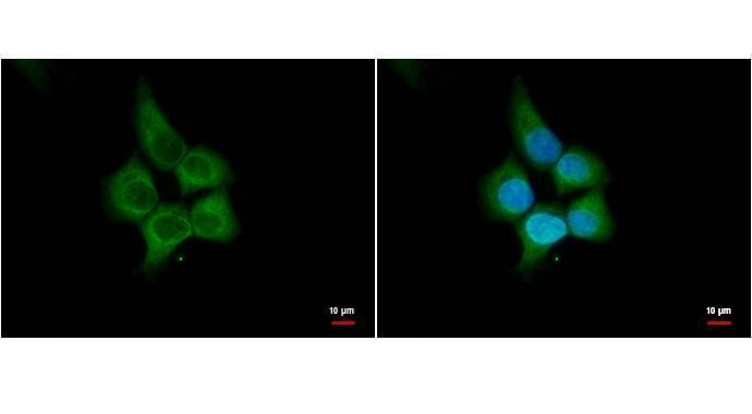
- HHIP antibody [N3C2], Internal antibody detects HHIP protein by immunofluorescent analysis. Sample: A431 cells were fixed in 4% paraformaldehyde for 15 min. Green: HHIP protein stained by HHIP antibody [N3C2], Internal antibody (GTX112584) diluted at 1:500. Blue: Hoechst 33342 staining. Scale bar = 10 £gm.
Supportive validation
- Submitted by
- GeneTex (provider)
- Main image

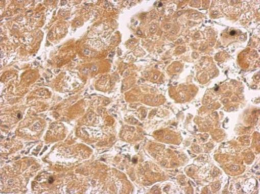
- Experimental details
- HHIP antibody [N3C2], Internal detects HHIP protein at cytosol on human hepatoma by immunohistochemical analysis. Sample: Paraffin-embedded hepatoma. HHIP antibody [N3C2], Internal (GTX112584) dilution: 1:500.