 Explore
 Validate
 Learn
Learn Western blot
 Immunohistochemistry
Antibody data
- Antibody Data
- Antigen structure
- References [4]
- Comments [0]
- Validations
- Immunohistochemistry [1]
Submit
Validation data
Reference
Comment
Report error
- Product number
- HPA031314 - Provider product page

- Provider
- Atlas Antibodies
- Proper citation
- Atlas Antibodies Cat#HPA031314, RRID:AB_10610278
- Product name
- Anti-PGD
- Antibody type
- Polyclonal
- Description
- Polyclonal Antibody against Human PGD, Gene description: phosphogluconate dehydrogenase, Validated applications: IHC, WB, Uniprot ID: P52209, Storage: Store at +4°C for short term storage. Long time storage is recommended at -20°C.
- Reactivity
- Human, Mouse
- Host
- Rabbit
- Conjugate
- Unconjugated
- Isotype
- IgG
- Vial size
- 100 µl
- Concentration
- 0.1 mg/ml
- Storage
- Store at +4°C for short term storage. Long time storage is recommended at -20°C.
- Handling
- The antibody solution should be gently mixed before use.
Submitted references Enzymatic Depletion of Mitochondrial Inorganic Polyphosphate (polyP) Increases the Generation of Reactive Oxygen Species (ROS) and the Activity of the Pentose Phosphate Pathway (PPP) in Mammalian Cells
Blockade of 6-phosphogluconate dehydrogenase generates CD8+ effector T cells with enhanced anti-tumor function
A feedback loop between the androgen receptor and 6-phosphogluoconate dehydrogenase (6PGD) drives prostate cancer growth
6-Phosphogluconate dehydrogenase (6PGD), a key checkpoint in reprogramming of regulatory T cells metabolism and function
Hambardikar V, Guitart-Mampel M, Scoma E, Urquiza P, Nagana G, Raftery D, Collins J, Solesio M
Antioxidants 2022;11(4):685
Antioxidants 2022;11(4):685
Blockade of 6-phosphogluconate dehydrogenase generates CD8+ effector T cells with enhanced anti-tumor function
Daneshmandi S, Cassel T, Lin P, Higashi R, Wulf G, Boussiotis V, Fan T, Seth P
Cell Reports 2021;34(10):108831
Cell Reports 2021;34(10):108831
A feedback loop between the androgen receptor and 6-phosphogluoconate dehydrogenase (6PGD) drives prostate cancer growth
Gillis J, Hinneh J, Ryan N, Irani S, Moldovan M, Quek L, Shrestha R, Hanson A, Xie J, Hoy A, Holst J, Centenera M, Mills I, Lynn D, Selth L, Butler L
eLife 2021;10
eLife 2021;10
6-Phosphogluconate dehydrogenase (6PGD), a key checkpoint in reprogramming of regulatory T cells metabolism and function
Daneshmandi S, Cassel T, Higashi R, Fan T, Seth P
eLife 2021;10
eLife 2021;10
No comments: Submit comment
Supportive validation
- Submitted by
- Atlas Antibodies (provider)
- Enhanced method
- Orthogonal validation
- Main image
- Experimental details
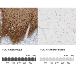
- Immunohistochemistry analysis in human esophagus and skeletal muscle tissues using Anti-PGD antibody. Corresponding PGD RNA-seq data are presented for the same tissues.
- Sample type
- Human
- Protocol
- Protocol
