Explore
 Validate
 Learn
Learn Immunohistochemistry
Antibody data
- Antibody Data
- Antigen structure
- References [1]
- Comments [0]
- Validations
- Immunohistochemistry [5]
- Other assay [1]
Submit
Validation data
Reference
Comment
Report error
- Product number
- PA5-59644 - Provider product page

- Provider
- Invitrogen Antibodies
- Product name
- SGLT2 Polyclonal Antibody
- Antibody type
- Polyclonal
- Antigen
- Recombinant protein fragment
- Description
- Immunogen sequence: FHEVGGYSGL FDKYLGAATS LTVSEDPAVG NISSFCYRPR PDSYHLL Highest antigen sequence identity to the following orthologs: Mouse - 89%, Rat - 91%.
- Reactivity
- Human
- Host
- Rabbit
- Isotype
- IgG
- Vial size
- 100 μL
- Concentration
- 0.10 mg/mL
- Storage
- Store at 4°C short term. For long term storage, store at -20°C, avoiding freeze/thaw cycles.
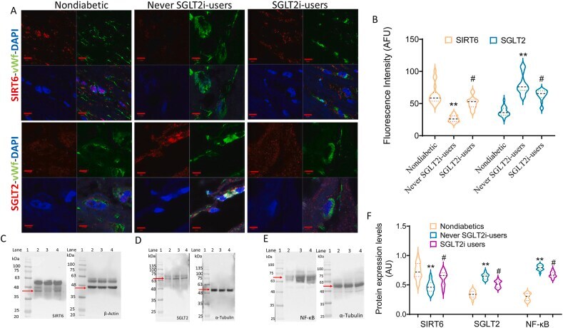
Submitted references Sodium-glucose co-transporter2 expression and inflammatory activity in diabetic atherosclerotic plaques: Effects of sodium-glucose co-transporter2 inhibitor treatment.
D'Onofrio N, Sardu C, Trotta MC, Scisciola L, Turriziani F, Ferraraccio F, Panarese I, Petrella L, Fanelli M, Modugno P, Massetti M, Marfella LV, Sasso FC, Rizzo MR, Barbieri M, Furbatto F, Minicucci F, Mauro C, Federici M, Balestrieri ML, Paolisso G, Marfella R
Molecular metabolism 2021 Dec;54:101337
Molecular metabolism 2021 Dec;54:101337
No comments: Submit comment
Supportive validation
- Submitted by
- Invitrogen Antibodies (provider)
- Main image
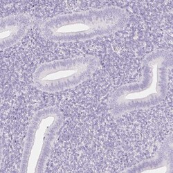
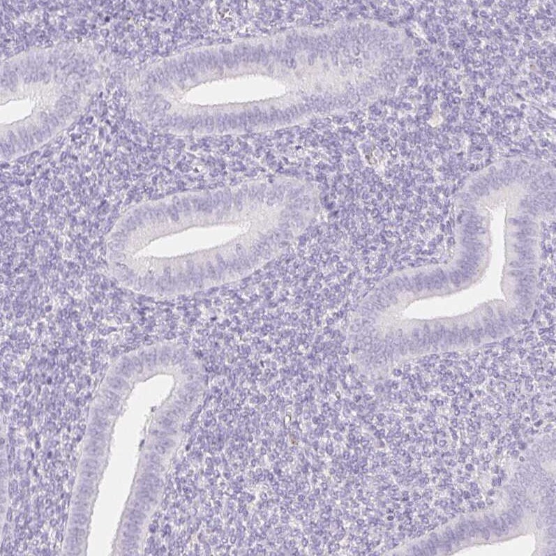
- Experimental details
- Immunohistochemical analysis of SGLT2 in human endometrium using SGLT2 Polyclonal Antibody (Product # PA5-59644) shows no positivity in glandular cells as expected.
- Submitted by
- Invitrogen Antibodies (provider)
- Main image
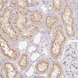
- Experimental details
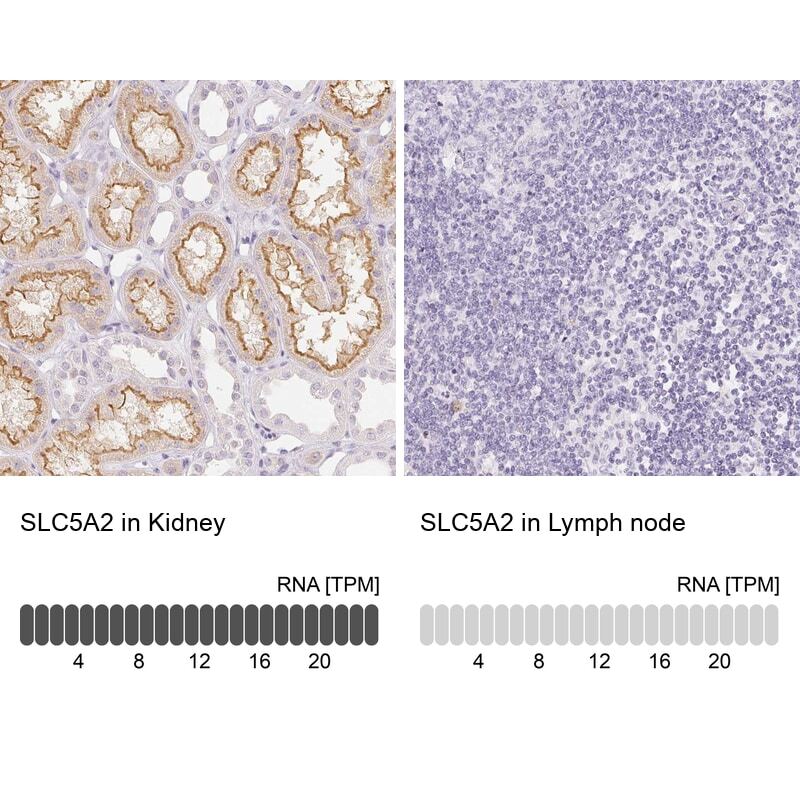
- Immunohistochemical analysis of SGLT2 in human kidney using SGLT2 Polyclonal Antibody (Product # PA5-59644) shows moderate positivity in apical membrane in cells in tubules.

- Submitted by
- Invitrogen Antibodies (provider)
- Main image
- Experimental details
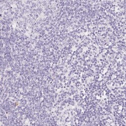
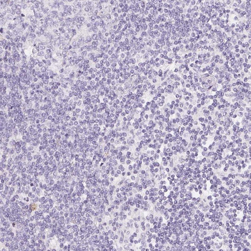
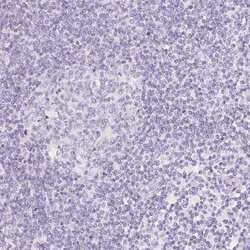
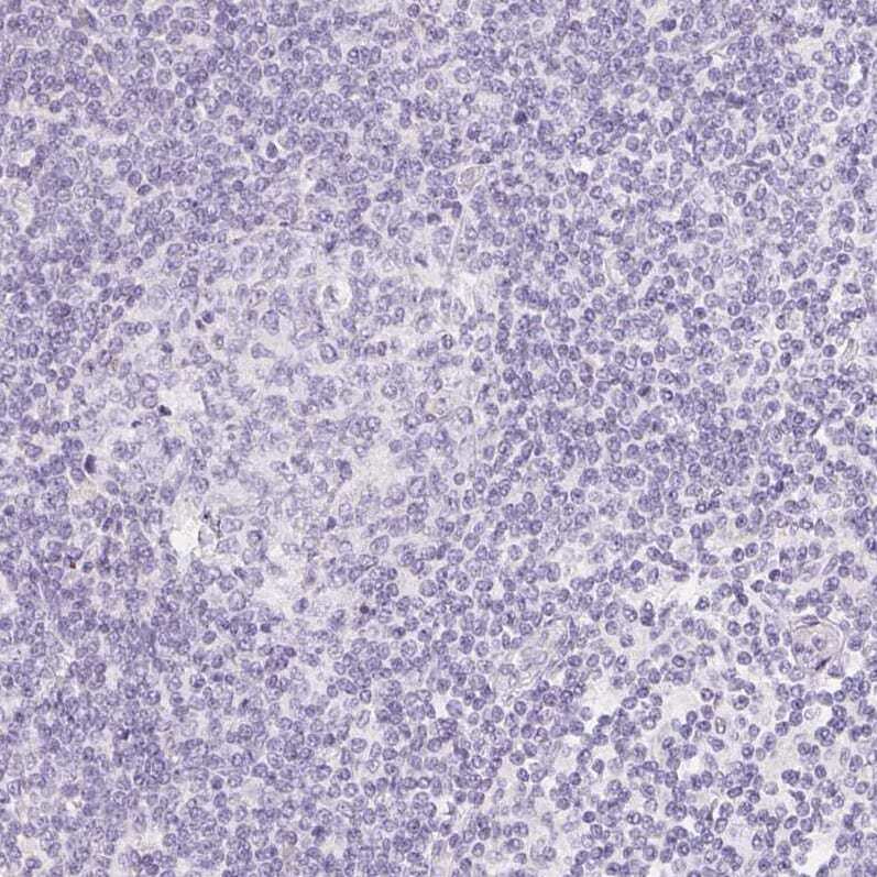
- Immunohistochemical staining of SGLT2 in human lymph node using SGLT2 Polyclonal Antibody (Product # PA5-59644) shows low expression as expected.
- Submitted by
- Invitrogen Antibodies (provider)
- Main image
- Experimental details
- Immunohistochemical analysis of SGLT2 in human lymph node using SGLT2 Polyclonal Antibody (Product # PA5-59644) shows no positivity in non-germinal center cells as expected.
- Submitted by
- Invitrogen Antibodies (provider)
- Main image
- Experimental details
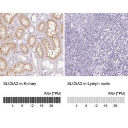
- Immunohistochemical staining of SGLT2 in human kidney and lymph node tissues using SGLT2 Polyclonal Antibody (Product # PA5-59644). Corresponding SLC5A2 RNA-seq data are presented for the same tissues.
Supportive validation
- Submitted by
- Invitrogen Antibodies (provider)
- Main image
- Experimental details
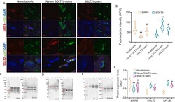
- Figure 1 SIRT6 and SGLT2 expression in atherosclerotic plaques . ( A ) Representative confocal laser-scanning microscope images of SIRT6 and SGLT2 expression levels (red) in deparaffinized atherosclerotic plaques from non-diabetic patients, current SGLT2i users, and never SGLTi users. The von Willebrand factor (vWf, green) was used to properly localize the immunofluorescence signals in endothelial cells, while DAPI staining was used for nuclei counterstaining (blue). Scale Bar = 5 mum. (B) The ImageJ software carried out arbitrary fluorescence units (AFU) of SIRT6 and SGLT2. **p < 0.01 versus plaque specimen from patients without diabetes, #p < 0.05 never SGLT2i users. Protein expression levels of (C) SIRT6 (D) SGLT2 and (E) NF- B ( NF - kappaB ) in plaques from diabetic, non-diabetic, and diabetic SGLT2i-user patients. (F) Protein quantification was performed using beta-Actin and alpha-tubulin as the internal control. SGLT2 protein expression detected by using anti-SGLT2 antibody from Cell Signaling Technology. Lane 1 = protein ladder molecular weight markers, lane 2 = non-diabetic, lane 3 = never SGLT2i users, lane 4 = SGLT2i users. The analysis of densitometric intensity was calculated with ImageJ software and expressed as arbitrary units (AU) with **p < 0.01 versus patients without diabetes, #p < 0.05 versus never SGLT2i users. Figure 1