Explore
 Validate
 Learn
Learn Western blot
 ELISA
ELISAAntibody data
- Antibody Data
- Antigen structure
- References [1]
- Comments [0]
- Validations
- Western blot [2]
- Immunocytochemistry [7]
- Immunoprecipitation [1]
- Immunohistochemistry [2]
- Other assay [5]
Submit
Validation data
Reference
Comment
Report error
- Product number
- PA5-116815 - Provider product page

- Provider
- Invitrogen Antibodies
- Product name
- GSDMD Polyclonal Antibody
- Antibody type
- Polyclonal
- Antigen
- Recombinant protein fragment
- Description
- Positive Samples: PC-3, THP-1, Jurkat Immunogen sequence: DGPAGAVLEC LVLSSGMLVP ELAIPVVYLL GALTMLSETQ HKLLAEALES QTLLGPLELV GSLLEQSAPW QERSTMSLPP GLLGNSWGEG APAWVLLDEC GLELGEDTPH VCWEPQAQGR MCALYASLAL LSGLSQEPH
- Reactivity
- Human, Mouse, Rat
- Host
- Rabbit
- Isotype
- IgG
- Vial size
- 100 μL
- Concentration
- 1.00 mg/mL
- Storage
- -20°C, Avoid Freeze/Thaw Cycles
Submitted references Long noncoding RNA X-inactive specific transcript regulates NLR family pyrin domain containing 3/caspase-1-mediated pyroptosis in diabetic nephropathy.
Xu J, Wang Q, Song YF, Xu XH, Zhu H, Chen PD, Ren YP
World journal of diabetes 2022 Apr 15;13(4):358-375
World journal of diabetes 2022 Apr 15;13(4):358-375
No comments: Submit comment
Supportive validation
- Submitted by
- Invitrogen Antibodies (provider)
- Main image
- Experimental details
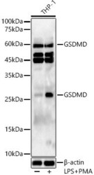
- Western blot analysis of GSDMD in various lysates. Samples were incubated with GSDMD Polyclonal antibody (Product # PA5-116815) using a dilution of 1:400, followed by HRP Goat Anti-Rabbit IgG (H+L) secondary antibody at a dilution of 1:10,000. THP-1 cells were treated by PMA/TPA (80 nM) at 37°C for overnight and LPS (1 µg/mL) at 37°C for 6 hours. Lysates/proteins: 25 µg per lane. Blocking buffer: 3% nonfat dry milk in TBST. Detection: ECL Basic Kit. Exposure time: 180s.

- Submitted by
- Invitrogen Antibodies (provider)
- Main image
- Experimental details
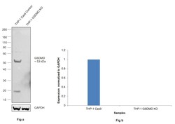
- Knockout of GSDMD was achieved by CRISPR-Cas9 genome editing using LentiArray™ Lentiviral sgRNA (Product # A32042, Assay ID CRISPR783871_LV) and LentiArray Cas9 Lentivirus (Product # A32064). Western blot analysis of GSDMD was performed by loading 30 µg of THP-1 Cas9 (Lane 1) and THP-1 GSDMD KO (Lane 2) whole cell extracts. The samples were electrophoresed using NuPAGE™ Novex™ 4-12% Bis-Tris Protein Gel (Product # NP0322BOX). Resolved proteins were then transferred onto a nitrocellulose membrane (Product # IB23001) by iBlot® 2 Dry Blotting System (Product # IB21001). The blot was probed with GSDMD Polyclonal Antibody (Product # PA5-116815, 1:1500 dilution) and detected by Goat anti-Rabbit IgG (H+L) Superclonal™ Recombinant Secondary Antibody, HRP (Product # A27036, 1:10,000 dilution) using the iBright™ FL1500 (Product # A44115). Chemiluminescent detection was performed using SuperSignal™ West Dura Extended Duration Substrate (Product # 34076). Loss of signal upon CRISPR mediated knockout (KO) using the LentiArray™ CRISPR product line confirms that antibody is specific to GSDMD. An uncharacterized band was observed at ~20 kDa in wild type sample lane.

Supportive validation
- Submitted by
- Invitrogen Antibodies (provider)
- Main image
- Experimental details
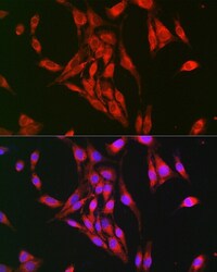
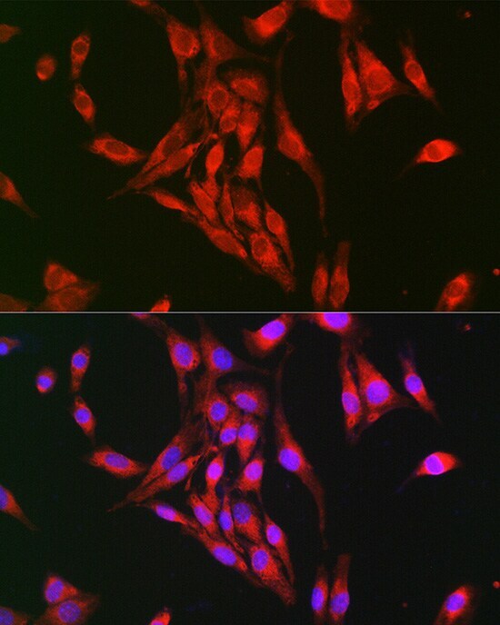
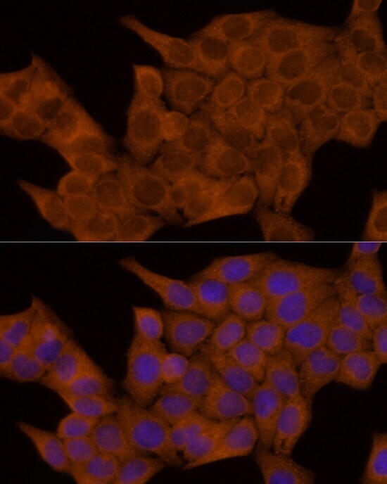
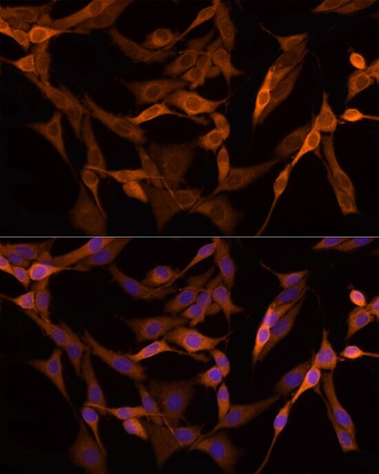
- Immunocytochemistry/Immunofluorescence analysis of GSDMD in BALB-3T3 cells using GSDMD Polyclonal Antibody (Product # PA5-116815) at a dilution of 1:100. Blue: DAPI for nuclear staining.
- Submitted by
- Invitrogen Antibodies (provider)
- Main image

- Experimental details
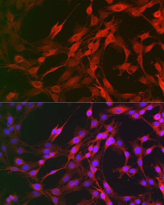
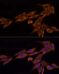
- Immunocytochemistry/Immunofluorescence analysis of GSDMD in C6 cells using GSDMD Polyclonal Antibody (Product # PA5-116815) at a dilution of 1:100. Blue: DAPI for nuclear staining.
- Submitted by
- Invitrogen Antibodies (provider)
- Main image
- Experimental details
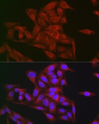
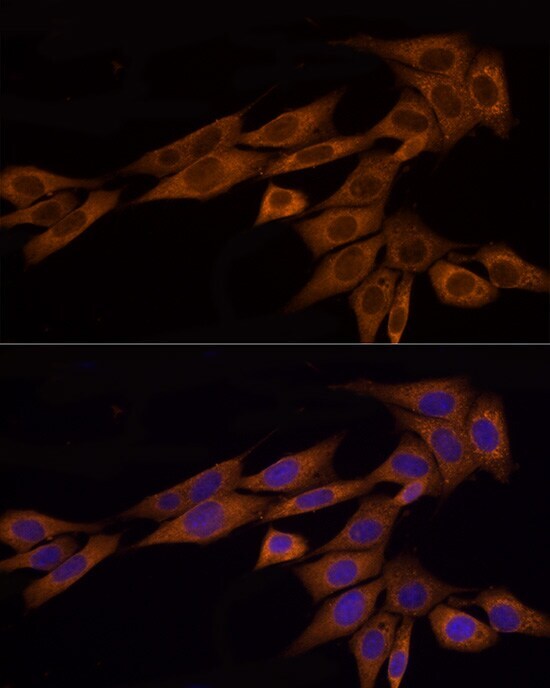
- Immunocytochemistry/Immunofluorescence analysis of GSDMD in U-2 OS cells using GSDMD Polyclonal Antibody (Product # PA5-116815) at a dilution of 1:100. Blue: DAPI for nuclear staining.
- Submitted by
- Invitrogen Antibodies (provider)
- Main image

- Experimental details
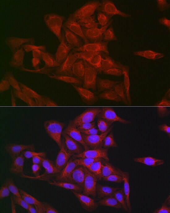
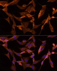
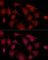
- Immunofluorescence analysis of GSDMD in HeLa cells. Samples were incubated with GSDMD Polyclonal antibody (Product # PA5-116815) using a dilution of 1:100 (40x lens). Blue: DAPI for nuclear staining.
- Submitted by
- Invitrogen Antibodies (provider)
- Main image
- Experimental details
- Immunofluorescence analysis of GSDMD in NIH/3T3 cells. Samples were incubated with GSDMD Polyclonal antibody (Product # PA5-116815) using a dilution of 1:100 (40x lens). Blue: DAPI for nuclear staining.
- Submitted by
- Invitrogen Antibodies (provider)
- Main image
- Experimental details
- Immunofluorescence analysis of GSDMD in PC-12 cells. Samples were incubated with GSDMD Polyclonal antibody (Product # PA5-116815) using a dilution of 1:100 (40x lens). Blue: DAPI for nuclear staining.
- Submitted by
- Invitrogen Antibodies (provider)
- Main image
- Experimental details
- Immunofluorescence analysis of GSDMD in PC-3 cells. Samples were incubated with GSDMD Polyclonal antibody (Product # PA5-116815) using a dilution of 1:100 (40x lens). Blue: DAPI for nuclear staining.

Supportive validation
- Submitted by
- Invitrogen Antibodies (provider)
- Main image

- Experimental details
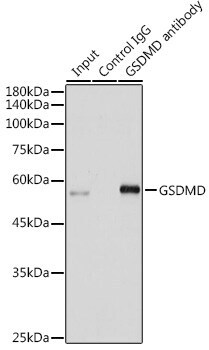
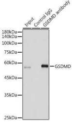
- Immunoprecipitation of GSDMD in 300 μg extracts of Jurkat cells. Samples were precipitated with 3 μg GSDMD Polyclonal antibody (Product # PA5-116815). Western blot was performed from the immunoprecipitate using GSDMD Polyclonal antibody (Product # PA5-116815) at a dilution of 1:500.
Supportive validation
- Submitted by
- Invitrogen Antibodies (provider)
- Main image

- Experimental details
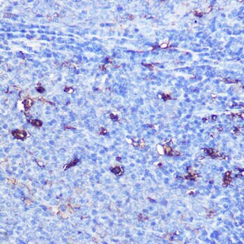
- Immunohistochemistry analysis of GSDMD in paraffin-embedded rat spleen. Samples were incubated with GSDMD Polyclonal antibody (Product # PA5-116815) using a dilution of 1:100 (40x lens). Perform microwave antigen retrieval with 10 mM Tris/EDTA buffer pH 9.0 before commencing with IHC staining protocol.

- Submitted by
- Invitrogen Antibodies (provider)
- Main image

- Experimental details
- Immunohistochemistry analysis of GSDMD in paraffin-embedded rat spleen. Samples were incubated with GSDMD Polyclonal antibody (Product # PA5-116815) using a dilution of 1:100 (40x lens). Perform microwave antigen retrieval with 10 mM Tris/EDTA buffer pH 9.0 before commencing with IHC staining protocol.
Supportive validation
- Submitted by
- Invitrogen Antibodies (provider)
- Main image
- Experimental details
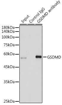
- Immunoprecipitation analysis of GSDMD in 300 µg extracts of Jurkat cells using GSDMD Polyclonal Antibody (Product # PA5-116815). Western blot was performed from the immunoprecipitate using the same antibody at a dilution of 1:500
- Submitted by
- Invitrogen Antibodies (provider)
- Main image

- Experimental details
- Immunoprecipitation analysis of GSDMD in 300 µg extracts of Jurkat cells using GSDMD Polyclonal Antibody (Product # PA5-116815). Western blot was performed from the immunoprecipitate using the same antibody at a dilution of 1:500

- Submitted by
- Invitrogen Antibodies (provider)
- Main image
- Experimental details
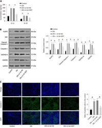
- Silencing lncRNA X inactive specific transcript can inhibit renal tubular epithelial cell pyroptosis in diabetic nephropathy rats. After establishing diabetic nephropathy model rats, lentivirus-LV sh- X inactive specific transcript was injected into the tail vein. A: Enzyme-linked immunosorbent assay detected IL-1beta and IL-18 expressions; B: Western blot tested the levels of NLR family pyrin domain containing 3, ASC, Cleaved Caspase-1, Caspase-1, GSDMD, and GSDMD-N; C: Immunofluorescence tested the expression of Caspase-1; scale bar: 25 mum; N = 8/group. The data were described as mean +- SD and analyzed by one-way ANOVA and Tukey's multiple comparisons test; a P < 0.05. DN: Diabetic nephropathy; LV: Lentivirus; XIST: X inactive specific transcript; sh: Short hairpin RNA; NLRP3: NLR family pyrin domain containing 3; ASC: Apoptosis speck-like protein; GSDMD: Gasdermin D; DAPI: 4',6-diamidino-2-phenylindole.

- Submitted by
- Invitrogen Antibodies (provider)
- Main image
- Experimental details
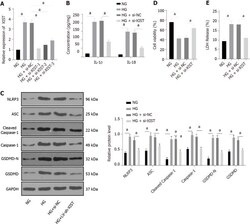
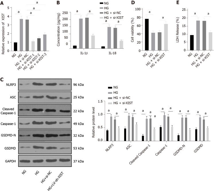
- Silencing X inactive specific transcript in diabetic nephropathy can inhibit renal tubular epithelial cell pyroptosis in high glucose-induced HK2 cells. si-NC or si- X inactive specific transcript (XIST) was delivered into HK2 cells treated with high glucose. A: The XIST expression detected by qRT-PCR; B: Enzyme-linked immunosorbent assay detected the expression of IL-1beta and IL-18; C: Western blot tested the levels of NLR family pyrin domain containing 3, ASC, Cleaved Caspase-1, Caspase-1, GSDMD, and GSDMD-N; D: Cell viability; E: Lactate dehydrogenase activity. The cell experiment was performed in triplicate. The data were described as mean +- SD and analyzed by one-way ANOVA (A/D-E) or two-way ANOVA (B/C) and Tukey's multiple comparisons test; a P < 0.05. NG: Normal glucose; HG: High glucose; si: Small interfering RNA; XIST: X inactive specific transcript; LDH: Lactate dehydrogenase; NLRP3: NLR family pyrin domain containing 3; SC: Apoptosis speck-like protein; GSDMD: Gasdermin D.
- Submitted by
- Invitrogen Antibodies (provider)
- Main image
- Experimental details
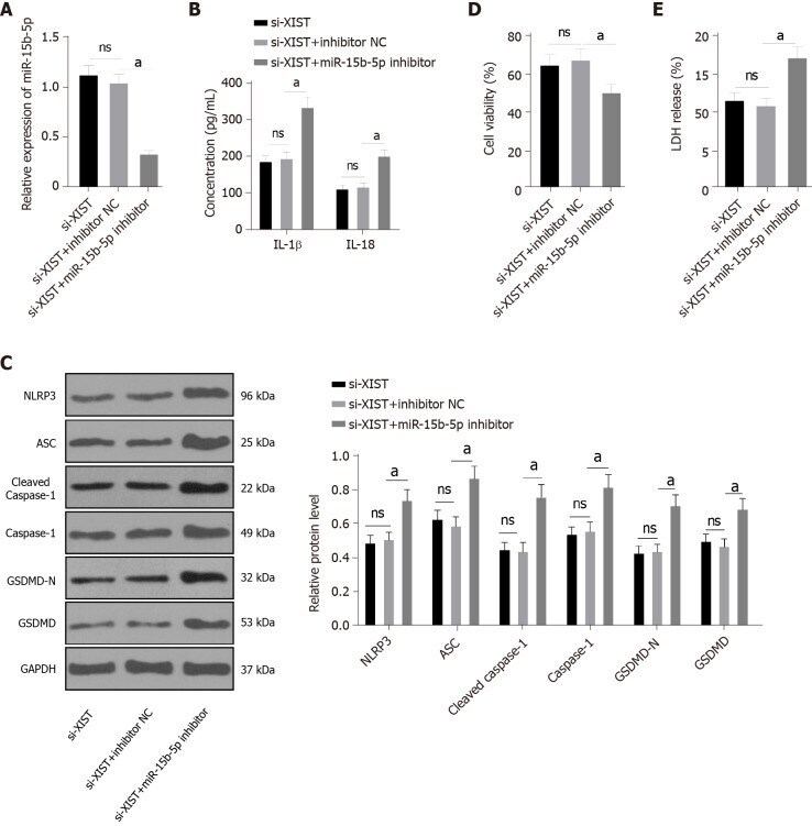
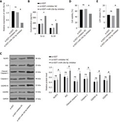
- Inhibition of miR-15b-5p in X inactive specific transcript -silenced HK2 cells activates renal tubular epithelial cell pyroptosis. In HG-treated HK2 cells, both X inactive specific transcript (XIST) and miR-15b-5p were silenced. A: qRT-PCR verified the transfection efficiency of miR-15b-5p inhibitor; B: Enzyme-linked immunosorbent assay detected the expression of IL-1beta and IL-18; C: Western blot tested the levels of NLR family pyrin domain containing 3, ASC, Cleaved Caspase-1, Caspase-1, GSDMD, and GSDMD-N; D: Cell viability; E: Lactate dehydrogenase activity. The cell experiment was performed in triplicate. The data were described as mean +- SD and analyzed by one-way ANOVA (A/D-E) or two-way ANOVA (B/C) and Tukey's multiple comparisons test; a P < 0.05. LDH: Lactate dehydrogenase; XIST: X inactive specific transcript; si: Small interfering RNA; NLRP3: NLR family pyrin domain containing 3; ASC: Apoptosis speck-like protein; GSDMD: Gasdermin D.