Explore
 Validate
 Learn
Learn Western blot
Antibody data
- Antibody Data
- Antigen structure
- References [0]
- Comments [0]
- Validations
- Western blot [1]
- Immunohistochemistry [1]
Submit
Validation data
Reference
Comment
Report error
- Product number
- AF4545 - Provider product page

- Provider
- Novus Biologicals
- Product name
- Goat Polyclonal MESDC2 Antibody
- Antibody type
- Polyclonal
- Description
- Immunogen affinity purified. Detects mouse MESDC2 in direct ELISAs and Western blots. In direct ELISAs, approximately 25% cross-reactivity with human MESDC2 is observed.
- Reactivity
- Mouse
- Host
- Goat
- Isotype
- IgG
- Vial size
- 100 ug
- Concentration
- LYOPH
- Storage
- Use a manual defrost freezer and avoid repeated freeze-thaw cycles. 12 months from date of receipt, -20 to -70 degreesC as supplied. 1 month, 2 to 8 degreesC under sterile conditions after reconstitution. 6 months, -20 to -70 degreesC under sterile conditions after reconstitution.
No comments: Submit comment
Supportive validation
- Submitted by
- Novus Biologicals (provider)
- Main image
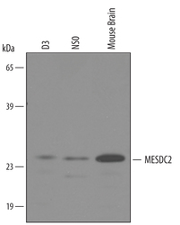
- Experimental details
- Detection of Mouse MESDC2 by Western Blot. Western blot shows lysates of mouse brain tissue, NS0 mouse myeloma cell line, and D3 mouse embryonic stem cell line. PVDF membrane was probed with 1 µg/mL of Goat Anti-Mouse MESDC2 Antigen Affinity-purified Polyclonal Antibody (Catalog # AF4545) followed by HRP-conjugated Anti-Goat IgG Secondary Antibody (Catalog # HAF019). A specific band was detected for MESDC2 at approximately 25 kDa (as indicated). This experiment was conducted under reducing conditions and using Immunoblot Buffer Group 8.

Supportive validation
- Submitted by
- Novus Biologicals (provider)
- Main image
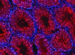
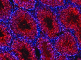
- Experimental details
- MESDC2 in Mouse Testis. MESDC2 was detected in perfusion fixed frozen sections of mouse testis using 10 µg/mL Goat Anti-Mouse MESDC2 Antigen Affinity-purified Polyclonal Antibody (Catalog # AF4545) overnight at 4 °C. Tissue was stained with the NorthernLights™ 557-conjugated Anti-Goat IgG Secondary Antibody (red; Catalog # NL001) and counterstained with DAPI (blue). View our protocol for Fluorescent IHC Staining of Frozen Tissue Sections.