Explore
 Validate
 Learn
Learn Western blot
 ELISA
ELISA Immunocytochemistry
 Immunohistochemistry
Antibody data
- Antibody Data
- Antigen structure
- References [0]
- Comments [0]
- Validations
- Western blot [2]
- Immunohistochemistry [3]
Submit
Validation data
Reference
Comment
Report error
- Product number
- LS-C745339 - Provider product page

- Provider
- LSBio
- Product name
- CASZ1 Antibody LS-C745339
- Antibody type
- Polyclonal
- Description
- Affinity purified
- Reactivity
- Human, Mouse, Drosophila
- Host
- Rabbit
- Isotype
- IgG
- Storage
- Store vial at -20°C or below prior to opening. Dilute 1:10 to minimize loss. Store the vial at -20°C or below after dilution. Avoid freeze-thaw cycles.
No comments: Submit comment
Enhanced validation
- Submitted by
- LSBio (provider)
- Enhanced method
- Genetic validation
- Main image
- Experimental details
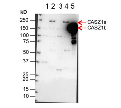
- Western Blot of Anti-CASZ1 Antibody. Lane 1: NBLS Cytoplasmic (20µg). Lane 2: NBLS Nuclear (3µg). Lane 3: BE2C Cytoplasmic (30µg). Lane 4: BE2C Nuclear (7µg). Lane 5: SY5Y-CASZ1b (10µg). Block: 5% Blotto/TTBS for 1 hour. Primary: Casz1 1:10,000 for 1 hour. Secondary: Goat anti-Rabbit HRP for 1 hour. 240sec exposure. Detects nuclear endogenous CASZ1a and CASZ1b; and transiently transfected CASZ1b isoform.

- Submitted by
- LSBio (provider)
- Enhanced method
- Genetic validation
- Main image

- Experimental details
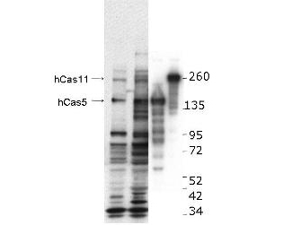
- Western blot using the anti-hCASZ1 antibody. This blot shows detection of endogenous and transfected human CASZ1 protein in fresh whole cell lysate (~30 µg). Protein was resolved by SDS-PAGE and transferred onto nitrocellulose. After blocking, the membrane was probed with the primary antibody diluted to 1:1,000, incubated 1.5 hours at room temperature, and incubated with HRP-conjugated Goat Anti-Rabbit antibody for 45 min. at room temperature. Lane 1, BE2(s) cell lysate. Lane 2, BE2(N) cell lysate. Lane 3, SY5Y transfected with hCas5 (125kDa). Lane 4, SY5Y transfected with hCas11 (190kDa).
Supportive validation
- Submitted by
- LSBio (provider)
- Enhanced method
- Genetic validation
- Main image

- Experimental details
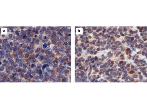
- Immunohistochemistry results of rabbit Anti-hCasz1 Antibody. Tissue: NB patient tumor. A. CASZ1 localized exclusively in the cytoplasm. B. CASZ1 localized in the cytoplasm and nucleus. Primary Antibody: Rabbit Anti-CASZ1 stained brown. Nucleus counterstained with hematoxylin (blue).

- Submitted by
- LSBio (provider)
- Main image

- Experimental details
- Immunohistochemistry results of rabbit Anti-hCasz1 Antibody. Tissue: NB patient tumor. A. CASZ1 localized exclusively in the cytoplasm. B. CASZ1 localized in the cytoplasm and nucleus. Primary Antibody: Rabbit Anti-CASZ1 stained brown. Nucleus counterstained with hematoxylin (blue).
- Submitted by
- LSBio (provider)
- Main image

- Experimental details
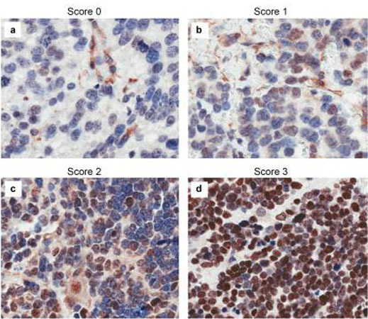
- Immunohistochemistry results of rabbit Anti-hCASZ1 Antibody. Tissue: NB patient tumor. A. Score 0- a rare positive nuclei. B. Score 1- (1-10% positive) equivocal/uninterpretable. C. Score 2- (10-50% positive) weak positive. D. Score 3- (>50% positive) strong positive. Primary Antibody: Rabbit Anti-CASZ1 stained brown. Nucleus counterstained with hematoxylin (blue). Localization: Nuclear.