Explore
 Validate
 Learn
Learn Western blot
 Immunohistochemistry
Antibody data
- Antibody Data
- Antigen structure
- References [4]
- Comments [0]
- Validations
- Western blot [2]
- Other assay [2]
Submit
Validation data
Reference
Comment
Report error
- Product number
- PA1-4656 - Provider product page

- Provider
- Invitrogen Antibodies
- Product name
- Dopamine Transporter Polyclonal Antibody
- Antibody type
- Polyclonal
- Antigen
- Synthetic peptide
- Description
- This antibody is specific for the ~88 kDa DAT protein in Western blots of SDS solubilized human and mouse striatal samples and in IHC applications with formaldehyde-fixed human and monkey (Macaque) brain sections.
- Reactivity
- Human, Mouse, Rat
- Host
- Rabbit
- Isotype
- IgG
- Vial size
- 100 µL
- Concentration
- 0.30 mg/mL
- Storage
- -20° C, Avoid Freeze/Thaw Cycles
Submitted references Single-cell transcriptomics of human iPSC differentiation dynamics reveal a core molecular network of Parkinson's disease.
Latent Tri-lineage Potential of Human Menstrual Blood-Derived Mesenchymal Stromal Cells Revealed by Specific In Vitro Culture Conditions.
Absence of Glia Maturation Factor Protects from Axonal Injury and Motor Behavioral Impairments after Traumatic Brain Injury.
Prolyl Oligopeptidase Regulates Dopamine Transporter Phosphorylation in the Nigrostriatal Pathway of Mouse.
Novak G, Kyriakis D, Grzyb K, Bernini M, Rodius S, Dittmar G, Finkbeiner S, Skupin A
Communications biology 2022 Jan 13;5(1):49
Communications biology 2022 Jan 13;5(1):49
Latent Tri-lineage Potential of Human Menstrual Blood-Derived Mesenchymal Stromal Cells Revealed by Specific In Vitro Culture Conditions.
Quintero-Espinosa D, Soto-Mercado V, Quintero-Quinchia C, Mendivil-Perez M, Velez-Pardo C, Jimenez-Del-Rio M
Molecular neurobiology 2021 Oct;58(10):5194-5209
Molecular neurobiology 2021 Oct;58(10):5194-5209
Absence of Glia Maturation Factor Protects from Axonal Injury and Motor Behavioral Impairments after Traumatic Brain Injury.
Selvakumar GP, Ahmed ME, Iyer SS, Thangavel R, Kempuraj D, Raikwar SP, Bazley K, Wu K, Khan A, Kukulka K, Bussinger B, Zaheer S, Burton C, James D, Zaheer A
Experimental neurobiology 2020 Jun 30;29(3):230-248
Experimental neurobiology 2020 Jun 30;29(3):230-248
Prolyl Oligopeptidase Regulates Dopamine Transporter Phosphorylation in the Nigrostriatal Pathway of Mouse.
Julku UH, Panhelainen AE, Tiilikainen SE, Svarcbahs R, Tammimäki AE, Piepponen TP, Savolainen MH, Myöhänen TT
Molecular neurobiology 2018 Jan;55(1):470-482
Molecular neurobiology 2018 Jan;55(1):470-482
No comments: Submit comment
Supportive validation
- Submitted by
- Invitrogen Antibodies (provider)
- Main image

- Experimental details
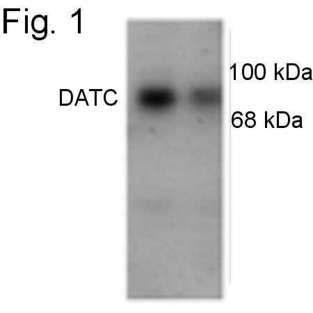
- Western blot of SDS-solubilized human caudate (60 µg and 20 µg protein per lane) showing the ~ 88 kDa band representing the DAT protein.
- Submitted by
- Invitrogen Antibodies (provider)
- Main image
- Experimental details
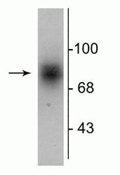
- Western blot of Dopamine Transporter in human striatal lysate showing specific immunolabeling of a band at ~88 kDa corresponding to Dopamine Transporter polyclonal antibody (Product # PA1-4656).

Supportive validation
- Submitted by
- Invitrogen Antibodies (provider)
- Main image
- Experimental details
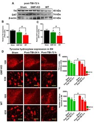
- Fig. 3 Removal of GMF improves TBI induced dopaminergic markers expression in SN of the midbrain. Induction of TBI by weight drop method in the cortical region causes significant reduction in dopaminergic markers such as TH and DAT expression as determined by western blot (A) and immunofluorescence (D; red fluorescence) in the SN region of the midbrain of WT and GMF-KO mice. Representative images show the TH-positive dopaminergic neurons in the SN at different magnifications (10X and 63X). Bar graphs show the quantitation of the western blot band intensity with TH (B), DAT (C) expression, TH-positive area (E; as arbitrary units) and the number of TH-positive dopaminergic neurons (F) as compared to the controls. Values are presented as mean+-SEM (n=6). *p

- Submitted by
- Invitrogen Antibodies (provider)
- Main image
- Experimental details
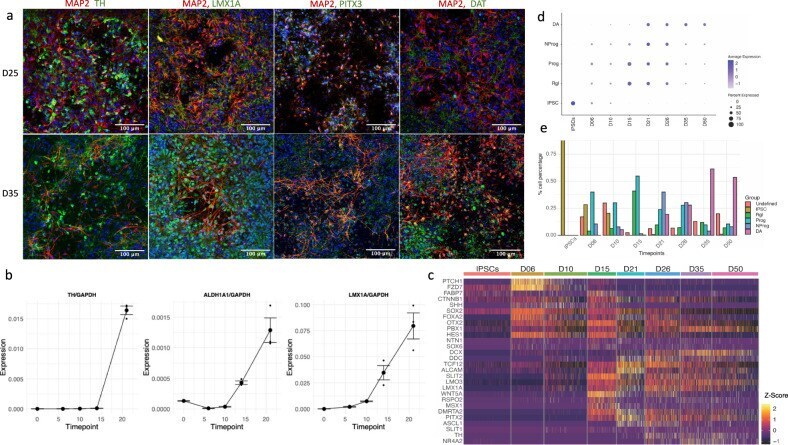
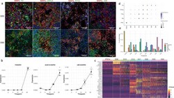
- Fig. 3 In vitro differentiation of iPSC-derived mDA neurons recapitulates the in vivo process. a To illustrate the maturation of neuronal morphology and mDA status, differentiated neurons were stained at D25 and D35 for a neuronal marker MAP2 (red) and mDA markers (green): TH, PITX3, LMX1A, and DAT. While D25 neurons show short processes and low expression of mDA markers, D35 neurons show much longer axons and well-defined expression of mDA markers (green/red overlap resulting on orange/yellow). b Quantitation of mDA markers TH, ALDH1A1 , and LMX1A , using absolute quantitation via qPCR. Each timepoint represents three independently differentiated biological replicates, amplified in duplicate. Standard error (SE) bars are the SE of biological replicates. The expression levels are standardized to total RNA and to the expression of the housekeeping gene GAPDH (see Methods). c Heatmap showing the expression of genes known from the literature to be involved and necessary for mDA neuron differentiation (Supplementary Table 3 ). Colors correlate to normalized counts ( z -score, centered, and scaled) of the indicated genes. d The mDA differentiation gene expression profile recently published by Asgrimsdottir and Arenas (2020) was used to show the progression during differentiation, from iPSCs to radial glia (Rgl), to progenitors (Prog) and neuroprogenitors (NProg), and to early mDA neurons (DA). Genes used to determine the expression modules are listed in Supplementary Table 3 . e P