Explore
 Validate
 Learn
Learn Western blot
Antibody data
- Antibody Data
- Antigen structure
- References [3]
- Comments [0]
- Validations
- Western blot [4]
- Immunocytochemistry [1]
- Immunohistochemistry [1]
Submit
Validation data
Reference
Comment
Report error
- Product number
- GTX108058 - Provider product page

- Provider
- GeneTex
- Proper citation
- GeneTex Cat#GTX108058, RRID:AB_1241448
- Product name
- VPS35 antibody [C3], C-term
- Antibody type
- Polyclonal
- Reactivity
- Human, Mouse, Rat
- Host
- Rabbit
Submitted references Phosphorylation of conserved phosphoinositide binding pocket regulates sorting nexin membrane targeting.
Proteomic analysis reveals novel common genes modulated in both replicative and stress-induced senescence.
VPS35 dysfunction impairs lysosomal degradation of α-synuclein and exacerbates neurotoxicity in a Drosophila model of Parkinson's disease.
Lenoir M, Ustunel C, Rajesh S, Kaur J, Moreau D, Gruenberg J, Overduin M
Nature communications 2018 Mar 8;9(1):993
Nature communications 2018 Mar 8;9(1):993
Proteomic analysis reveals novel common genes modulated in both replicative and stress-induced senescence.
Succoio M, Comegna M, D'Ambrosio C, Scaloni A, Cimino F, Faraonio R
Journal of proteomics 2015 Oct 14;128:18-29
Journal of proteomics 2015 Oct 14;128:18-29
VPS35 dysfunction impairs lysosomal degradation of α-synuclein and exacerbates neurotoxicity in a Drosophila model of Parkinson's disease.
Miura E, Hasegawa T, Konno M, Suzuki M, Sugeno N, Fujikake N, Geisler S, Tabuchi M, Oshima R, Kikuchi A, Baba T, Wada K, Nagai Y, Takeda A, Aoki M
Neurobiology of disease 2014 Nov;71:1-13
Neurobiology of disease 2014 Nov;71:1-13
No comments: Submit comment
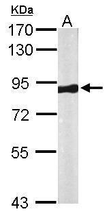
Supportive validation
- Submitted by
- GeneTex (provider)
- Main image
- Experimental details
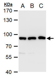
- Sample (50 ?g of whole cell lysate) A: Mouse brain 7.5% SDS PAGE GTX108058 diluted at 1:1000 The HRP-conjugated anti-rabbit IgG antibody (GTX213110-01) was used to detect the primary antibody.
- Submitted by
- GeneTex (provider)
- Main image
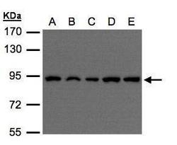
- Experimental details
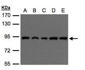
- Sample(30 ?g of whole cell lysate)A:H1299B:HeLa S3 (GTX14654)C:Hep G2 (GTX27900)D:MOLT4 (GTX27912)E:Raji (GTX27908)7.5% SDS PAGEGTX108058 diluted at 1:500The HRP-conjugated anti-rabbit IgG antibody (GTX213110-01) was used to detect the primary antibody.
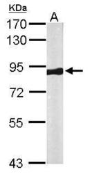
- Submitted by
- GeneTex (provider)
- Main image
- Experimental details
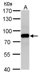
- VPS35 antibody detects VPS35 protein by western blot analysis.A. 50 ?g rat brain lysate/extract7.5% SDS-PAGEVPS35 antibody (GTX108058) dilution: 1:500 The HRP-conjugated anti-rabbit IgG antibody (GTX213110-01) was used to detect the primary antibody.

- Submitted by
- GeneTex (provider)
- Main image
- Experimental details
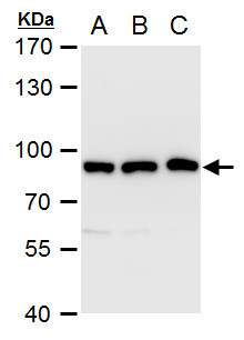
- VPS35 antibody [C3], C-term detects VPS35 protein by western blot analysis.A. 30 ?g A549 whole cell extract B. 30 ?g H1299 whole cell extract C. 30 ?g HCT116 whole cell extract7.5% SDS-PAGEVPS35 antibody [C3], C-term (GTX108058) dilution: 1:1000 The HRP-conjugated anti-rabbit IgG antibody (GTX213110-01) was used to detect the primary antibody.
Supportive validation
- Submitted by
- GeneTex (provider)
- Main image
- Experimental details
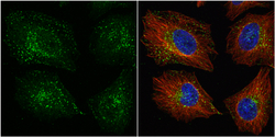
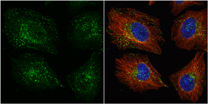
- VPS35 antibody [C3], C-term detects VPS35 protein at cytoplasm by immunofluorescent analysis.Sample: HeLa cells were fixed in 4% paraformaldehyde at RT for 15 min.Green: VPS35 protein stained by VPS35 antibody [C3], C-term (GTX108058) diluted at 1:1000.Red: alpha Tubulin, a cytoskeleton marker, stained by alpha Tubulin antibody [B-5-1-2] (GTX11304) diluted at 1:10000.Blue: Hoechst 33342 staining.
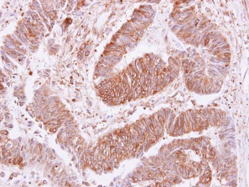
Supportive validation
- Submitted by
- GeneTex (provider)
- Main image

- Experimental details
- VPS35 antibody [C3], C-term detects VPS35 protein at cytoplasm and membrane on human colon carcinoma by immunohistochemical analysis. Sample: Paraffin-embedded colon carcinoma. VPS35 antibody [C3], C-term (GTX108058) dilution: 1:250.