Explore
 Validate
 Learn
Learn Western blot
 Immunocytochemistry
Antibody data
- Antibody Data
- Antigen structure
- References [2]
- Comments [0]
- Validations
- Western blot [1]
- Immunocytochemistry [1]
Submit
Validation data
Reference
Comment
Report error
- Product number
- HPA040802 - Provider product page

- Provider
- Atlas Antibodies
- Proper citation
- Atlas Antibodies Cat#HPA040802, RRID:AB_2677142
- Product name
- Anti-VPS35
- Antibody type
- Polyclonal
- Description
- Polyclonal Antibody against Human VPS35, Gene description: vacuolar protein sorting 35 homolog (S. cerevisiae), Alternative Gene Names: FLJ10752, MEM3, PARK17, Validated applications: ICC, WB, Uniprot ID: Q96QK1, Storage: Store at +4°C for short term storage. Long time storage is recommended at -20°C.
- Reactivity
- Human, Mouse, Rat
- Host
- Rabbit
- Conjugate
- Unconjugated
- Isotype
- IgG
- Vial size
- 100 µl
- Concentration
- 0.1 mg/ml
- Storage
- Store at +4°C for short term storage. Long time storage is recommended at -20°C.
- Handling
- The antibody solution should be gently mixed before use.
Submitted references SNX27-Mediated Recycling of Neuroligin-2 Regulates Inhibitory Signaling.
Identification of TMEM230 mutations in familial Parkinson's disease
Halff EF, Szulc BR, Lesept F, Kittler JT
Cell reports 2019 Nov 26;29(9):2599-2607.e6
Cell reports 2019 Nov 26;29(9):2599-2607.e6
Identification of TMEM230 mutations in familial Parkinson's disease
Deng H, Shi Y, Yang Y, Ahmeti K, Miller N, Huang C, Cheng L, Zhai H, Deng S, Nuytemans K, Corbett N, Kim M, Deng H, Tang B, Yang Z, Xu Y, Chan P, Huang B, Gao X, Song Z, Liu Z, Fecto F, Siddique N, Foroud T, Jankovic J, Ghetti B, Nicholson D, Krainc D, Melen O, Vance J, Pericak-Vance M, Ma Y, Rajput A, Siddique T
Nature Genetics 2016;48(7):733-739
Nature Genetics 2016;48(7):733-739
No comments: Submit comment
Enhanced validation
- Submitted by
- Atlas Antibodies (provider)
- Enhanced method
- Genetic validation
- Main image

- Experimental details
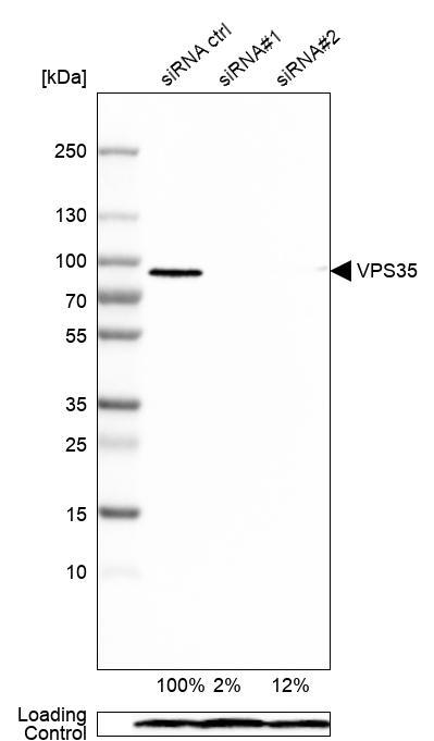
- Western blot analysis in HEK293 cells transfected with control siRNA, target specific siRNA probe #1 and #2, using Anti-VPS35 antibody. Remaining relative intensity is presented. Loading control: Anti-PPIB.
- Sample type
- Human
- Protocol
- Protocol
Supportive validation
- Submitted by
- Atlas Antibodies (provider)
- Main image
- Experimental details
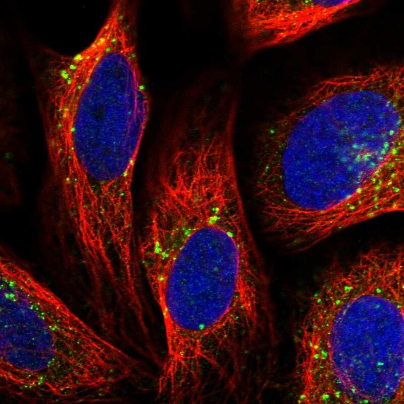
- Immunofluorescent staining of human cell line U-2 OS shows localization to endosomes & lysosomes.
- Sample type
- Human