 Explore
 Validate
 Learn
Learn Western blot
 Immunoprecipitation
Antibody data
- Antibody Data
- Antigen structure
- References [0]
- Comments [0]
- Validations
- Western blot [1]
- Immunocytochemistry [1]
Submit
Validation data
Reference
Comment
Report error
- Product number
- PA1-24970 - Provider product page

- Provider
- Invitrogen Antibodies
- Product name
- GAP43 Polyclonal Antibody
- Antibody type
- Polyclonal
- Antigen
- Purifed from natural sources
- Description
- PA1-24970 detects GAP-43 from human, mouse, rat, feline, and monkey samples.
- Reactivity
- Human, Mouse, Rat, Feline
- Host
- Rabbit
- Isotype
- IgG
- Vial size
- 50 µL
- Concentration
- Conc. Not Determined
- Storage
- Store at 4°C short term. For long term storage, store at -20°C, avoiding freeze/thaw cycles.
No comments: Submit comment
Supportive validation
- Submitted by
- Invitrogen Antibodies (provider)
- Main image

- Experimental details
- Western blot was performed using Anti-GAP43 Polyclonal Antibody (Product # PA1-24970) and a 38-48 kDa band corresponding to GAP43 was observed across cell lines and tissues tested except in Mouse Lung which is reported to be low to negative for GAP43 expression. An increase in GAP43 expression was observed in SH-SY5Y on neuronal differentiation. Membrane enriched extracts (30 µg lysate) of SH-SY5Y (Lane 1), SH-SY5Y differentiated to neurons (Lane 2), SK-N-SH (Lane 3), IMR-32 (Lane 4), Mouse Lung (Lane 5), Mouse Brain (Lane 6) and Rat Brain (Lane 7) were electrophoresed using NuPAGE™ 4-12% Bis-Tris Protein Gel (Product # NP0322BOX). Resolved proteins were then transferred onto a Nitrocellulose membrane (Product # IB23002) by iBlot® 2 Dry Blotting System (Product # IB21001). The blot was probed with the primary antibody (1:1000) and detected by chemiluminescence with Goat anti-Rabbit IgG (H+L) Superclonal™ Recombinant Secondary Antibody, HRP (Product # A27036,1:4000 dilution) using the iBright FL 1000 (Product # A32752). Chemiluminescent detection was performed using Novex® ECL Chemiluminescent Substrate Reagent Kit (Product # WP20005).

Supportive validation
- Submitted by
- Invitrogen Antibodies (provider)
- Main image
- Experimental details
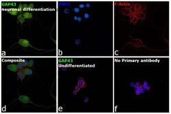
- Immunofluorescence analysis of GAP43 was performed using 70% confluent log phase PC-12 neuronal differentiated and undifferentiated cells. The cells were fixed with 4% paraformaldehyde for 10 minutes, permeabilized with 0.1% Triton™ X-100 for 15 minutes, and blocked with 2% BSA for 45 minutes at room temperature. The cells were labeled with GAP43 Polyclonal Antibody (Product # PA1-24970) at 1:100 dilution in 0.1% BSA, incubated at 4 degree celsius overnight and then labeled with Donkey anti-Rabbit IgG (H+L) Highly Cross-Adsorbed Secondary Antibody, Alexa Fluor Plus 488 (Product # A32790), (1:2000 dilution), for 45 minutes at room temperature (Panel a: Green). Nuclei (Panel b: Blue) were stained with ProLong™ Diamond Antifade Mountant with DAPI (Product # P36962). F-actin (Panel c: Red) was stained with Rhodamine Phalloidin (Product # R415, 1:300). Panel d represents the merged image showing Plasma membrane and cytoplasm localization in neuronal differentiated PC-12 cells, but not in undifferentiated PC-12 cells (Panel e). Panel f represents control cells with no primary antibody to assess the background. The images were captured at 60X magnification.
