Explore
 Validate
 Learn
Learn Western blot
Antibody data
- Antibody Data
- Antigen structure
- References [1]
- Comments [0]
- Validations
- Western blot [7]
- Immunocytochemistry [2]
- Immunohistochemistry [2]
Submit
Validation data
Reference
Comment
Report error
- Product number
- GTX127937 - Provider product page

- Provider
- GeneTex
- Product name
- GAP43 antibody
- Antibody type
- Polyclonal
- Reactivity
- Human, Mouse, Rat
- Host
- Rabbit
Submitted references Neuropathic pain-induced depressive-like behavior and hippocampal neurogenesis and plasticity are dependent on TNFR1 signaling.
Dellarole A, Morton P, Brambilla R, Walters W, Summers S, Bernardes D, Grilli M, Bethea JR
Brain, behavior, and immunity 2014 Oct;41:65-81
Brain, behavior, and immunity 2014 Oct;41:65-81
No comments: Submit comment
Supportive validation
- Submitted by
- GeneTex (provider)
- Main image

- Experimental details
- GAP43 antibody detects Gap43 protein by western blot analysis.A. 30 ?g SK-N-SH whole cell lysate/extractB. 30 ?g IMR32 whole cell lysate/extractC. 30 ?g SK-N-AS whole cell lysate/extract12% SDS-PAGEGAP43 antibody (GTX127937) dilution: 1:10000 The HRP-conjugated anti-rabbit IgG antibody (GTX213110-01) was used to detect the primary antibody.

- Submitted by
- GeneTex (provider)
- Main image

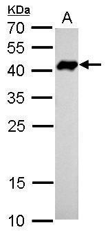
- Experimental details
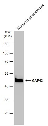
- GAP43 antibody detects Gap43 protein by western blot analysis.A. 5 ?g mouse brain lysate/extract12% SDS-PAGEGAP43 antibody (GTX127937) dilution: 1:5000 The HRP-conjugated anti-rabbit IgG antibody (GTX213110-01) was used to detect the primary antibody.
- Submitted by
- GeneTex (provider)
- Main image

- Experimental details
- GAP43 antibody detects Gap43 protein by western blot analysis.A. 1 ?g Rat brain lysate/extract12% SDS-PAGEGAP43 antibody (GTX127937) dilution: 1:5000 The HRP-conjugated anti-rabbit IgG antibody (GTX213110-01) was used to detect the primary antibody.

- Submitted by
- GeneTex (provider)
- Main image
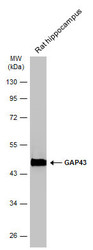
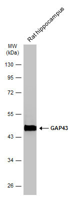
- Experimental details
- Rat tissue extract (50 ?g) was separated by 10% SDS-PAGE, and the membrane was blotted with GAP43 antibody (GTX127937) diluted at 1:30000. The HRP-conjugated anti-rabbit IgG antibody (GTX213110-01) was used to detect the primary antibody.
- Submitted by
- GeneTex (provider)
- Main image

- Experimental details
- Mouse tissue extract (50 ?g) was separated by 10% SDS-PAGE, and the membrane was blotted with GAP43 antibody (GTX127937) diluted at 1:30000. The HRP-conjugated anti-rabbit IgG antibody (GTX213110-01) was used to detect the primary antibody.
- Submitted by
- GeneTex (provider)
- Main image

- Experimental details
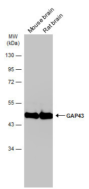
- Various tissue extracts (50 ?g) were separated by 10% SDS-PAGE, and the membrane was blotted with GAP43 antibody (GTX127937) diluted at 1:30000. The HRP-conjugated anti-rabbit IgG antibody (GTX213110-01) was used to detect the primary antibody.
Supportive validation
- Submitted by
- GeneTex (provider)
- Main image

- Experimental details
- GAP43 antibody detects GAP43 protein at membrane by immunofluorescent analysis.Sample: SKNSH cells were fixed in 4% paraformaldehyde at RT for 15 min.Green: GAP43 protein stained by GAP43 antibody (GTX127937) diluted at 1:500.Blue: Hoechst 33342 staining.

- Submitted by
- GeneTex (provider)
- Main image

- Experimental details
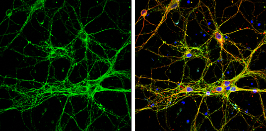
- GAP43 antibody detects GAP43 protein by immunofluorescent analysis.Sample: DIV9 rat E18 primary cortical neurons were fixed in 4% paraformaldehyde at RT for 15 min.Green: GAP43 protein stained by GAP43 antibody (GTX127937) diluted at 1:500.Red: beta Tubulin 3/ Tuj1, stained by beta Tubulin 3/ Tuj1 antibody [GT886] (GTX631830) diluted at 1:500.Blue: Fluoroshield with DAPI (GTX30920).
Supportive validation
- Submitted by
- GeneTex (provider)
- Main image
- Experimental details
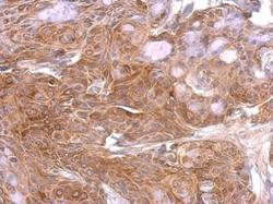
- GAP43 antibody detects Gap43 protein at on Cal27 xenograft by immunohistochemical analysis. Sample: Paraffin-embedded Cal27 xenograft. GAP43 antibody (GTX127937) dilution: 1:500.

- Submitted by
- GeneTex (provider)
- Main image

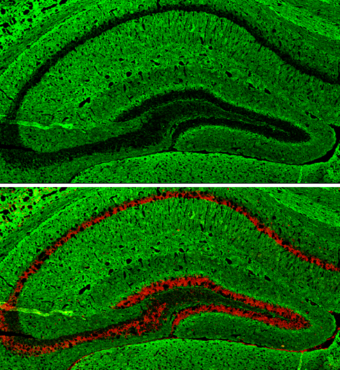
- Experimental details
- GAP43 antibody detects GAP43 protein expression by immunohistochemical analysis.Sample: Frozen-sectioned adult mouse hippocampus. Green: GAP43 protein stained by GAP43 antibody (GTX127937) diluted at 1:250.Red: NeuN, stained by NeuN antibody [2Q158] (GTX30773) diluted at 1:500.