Explore
 Validate
 Learn
Learn Western blot
 Immunocytochemistry
Antibody data
- Antibody Data
- Antigen structure
- References [1]
- Comments [0]
- Validations
- Immunocytochemistry [2]
- Immunohistochemistry [3]
- Other assay [1]
Submit
Validation data
Reference
Comment
Report error
- Product number
- PA5-79299 - Provider product page

- Provider
- Invitrogen Antibodies
- Product name
- GAP43 Polyclonal Antibody
- Antibody type
- Polyclonal
- Antigen
- Synthetic peptide
- Description
- Reconstitute with 0.2 mL of distilled water to yield a concentration of 500 µg/mL. Positive Control - WB: U87 whole cell, Rat Brain Tissue, Mouse Brain Tissue. IHC: Rat Brain Tissue, Human Meningeoma Tissue, Human Glioma Tissue.
- Reactivity
- Human, Mouse, Rat
- Host
- Rabbit
- Isotype
- IgG
- Vial size
- 100 μg
- Concentration
- 500 μg/mL
- Storage
- -20°C
Submitted references Temporal expression profiling of DAMPs-related genes revealed the biphasic post-ischemic inflammation in the experimental stroke model.
Yamaguchi A, Jitsuishi T, Hozumi T, Iwanami J, Kitajo K, Yamaguchi H, Mori Y, Mogi M, Sawai S
Molecular brain 2020 Apr 7;13(1):57
Molecular brain 2020 Apr 7;13(1):57
No comments: Submit comment
Supportive validation
- Submitted by
- Invitrogen Antibodies (provider)
- Main image
- Experimental details
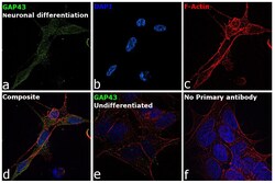
- Immunofluorescence analysis of GAP43 was performed using 70% confluent log phase SH-SY5Y cells differentiated to neurons and undifferentiated cells. The cells were fixed with 4% paraformaldehyde for 10 minutes, permeabilized with 0.1% Triton™ X-100 for 15 minutes, and blocked with 2% BSA for 45 minutes at room temperature. The cells were labeled with GAP43 Polyclonal Antibody (Product # PA5-79299) at 5 µg/mL in 0.1% BSA, incubated at 4-degree Celsius overnight and then labeled with Donkey anti-Rabbit IgG (H+L) Highly Cross-Adsorbed Secondary Antibody, Alexa Fluor Plus 488 (Product # A32790), (1:2000 dilution), for 45 minutes at room temperature (Panel a: Green). Nuclei (Panel b: Blue) were stained with ProLong™ Diamond Antifade Mountant with DAPI (Product # P36962). F-actin (Panel c: Red) was stained with Rhodamine Phalloidin (Product # R415, 1:300). Panel d represents the merged image showing Plasma membrane and cytoplasmic localization of the neuronal differentiated SH-SY5Y cells. Panel e represents undifferentiated SH-SY5Y cells with no expression for GAP43. Panel f represents control cells with no primary antibody to assess the background. The images were captured at 60X magnification.
- Submitted by
- Invitrogen Antibodies (provider)
- Main image

- Experimental details
- Immunofluorescence analysis of GAP43 was performed using 70% confluent log phase SH-SY5Y cells differentiated to neurons and undifferentiated cells. The cells were fixed with 4% paraformaldehyde for 10 minutes, permeabilized with 0.1% Triton™ X-100 for 15 minutes, and blocked with 2% BSA for 45 minutes at room temperature. The cells were labeled with GAP43 Polyclonal Antibody (Product # PA5-79299) at 5 µg/mL in 0.1% BSA, incubated at 4-degree Celsius overnight and then labeled with Donkey anti-Rabbit IgG (H+L) Highly Cross-Adsorbed Secondary Antibody, Alexa Fluor Plus 488 (Product # A32790), (1:2000 dilution), for 45 minutes at room temperature (Panel a: Green). Nuclei (Panel b: Blue) were stained with ProLong™ Diamond Antifade Mountant with DAPI (Product # P36962). F-actin (Panel c: Red) was stained with Rhodamine Phalloidin (Product # R415, 1:300). Panel d represents the merged image showing Plasma membrane and cytoplasmic localization of the neuronal differentiated SH-SY5Y cells. Panel e represents undifferentiated SH-SY5Y cells with no expression for GAP43. Panel f represents control cells with no primary antibody to assess the background. The images were captured at 60X magnification.

Supportive validation
- Submitted by
- Invitrogen Antibodies (provider)
- Main image
- Experimental details
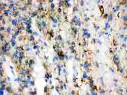
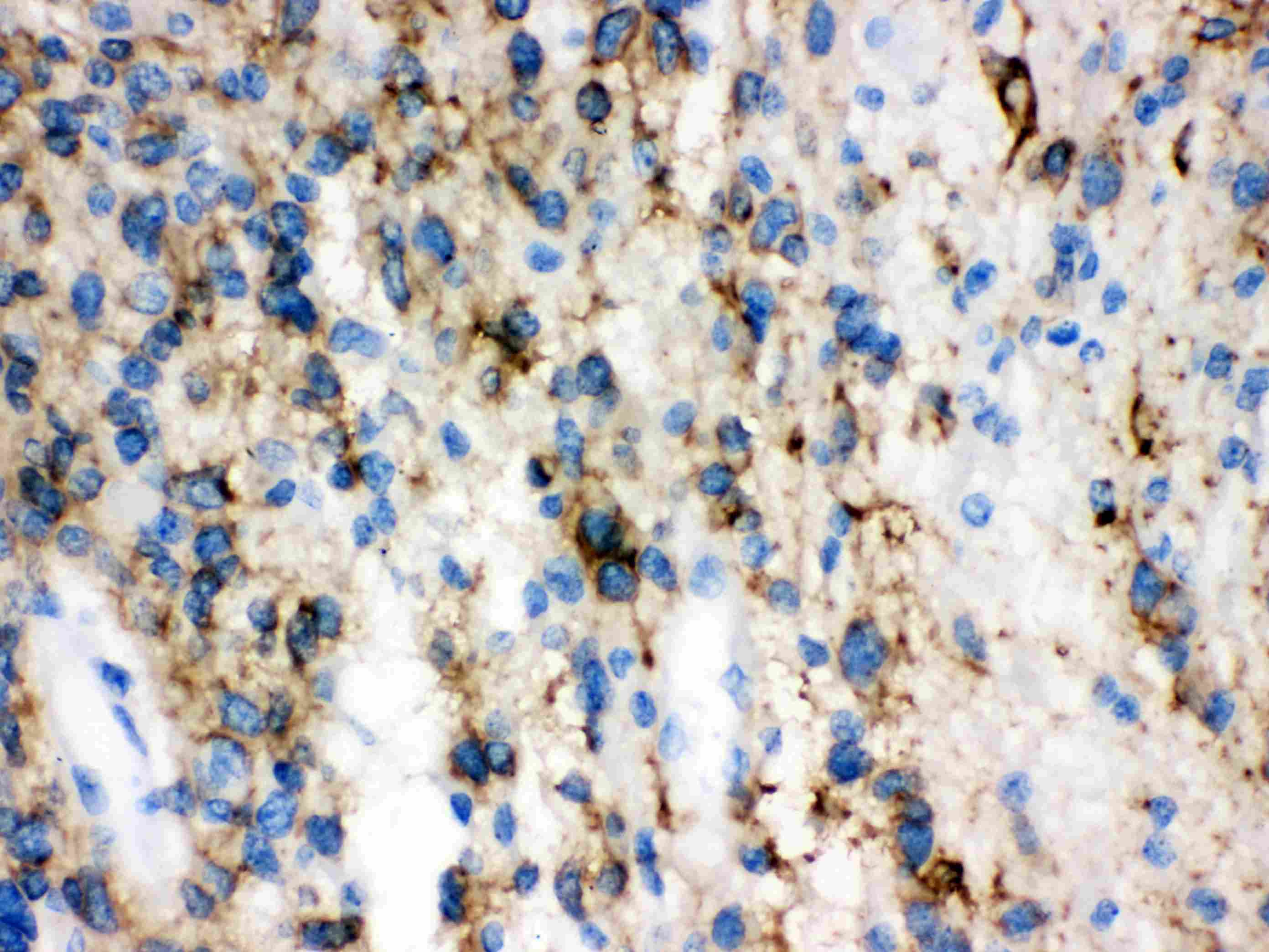
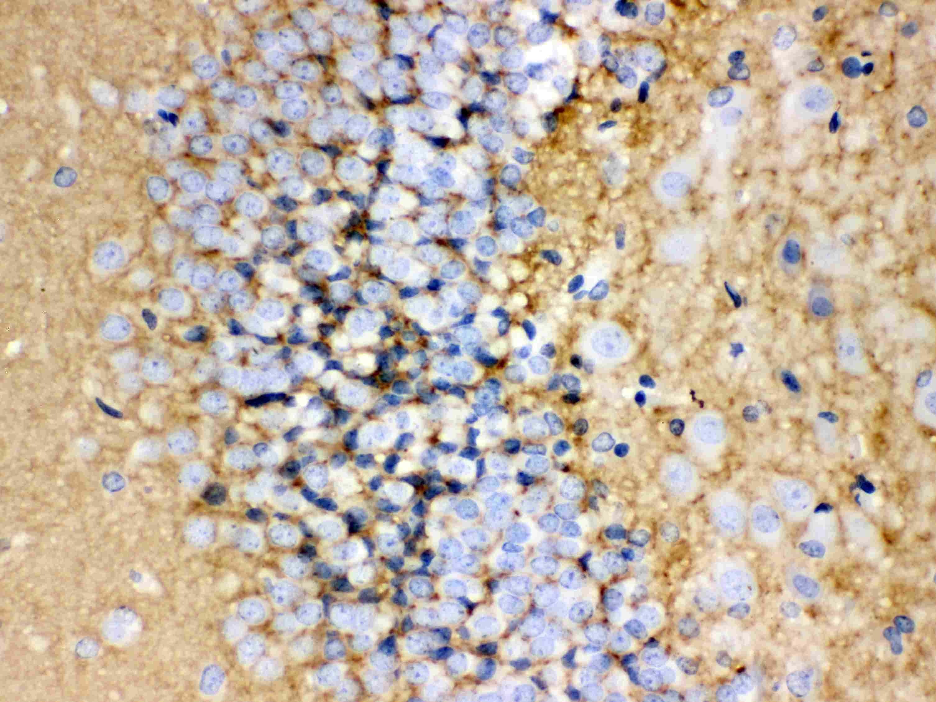
- Immunohistochemistry analysis of GAP43 on paraffin-embedded human glioma tissue. Sample was incubated with GAP43 polyclonal antibody (Product# PA5-79299).
- Submitted by
- Invitrogen Antibodies (provider)
- Main image
- Experimental details
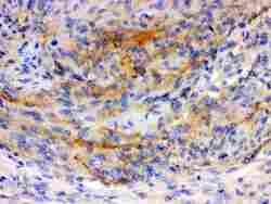
- Immunohistochemistry analysis of GAP43 on paraffin-embedded human meningioma tissue. Sample was incubated with GAP43 polyclonal antibody (Product# PA5-79299).
- Submitted by
- Invitrogen Antibodies (provider)
- Main image
- Experimental details
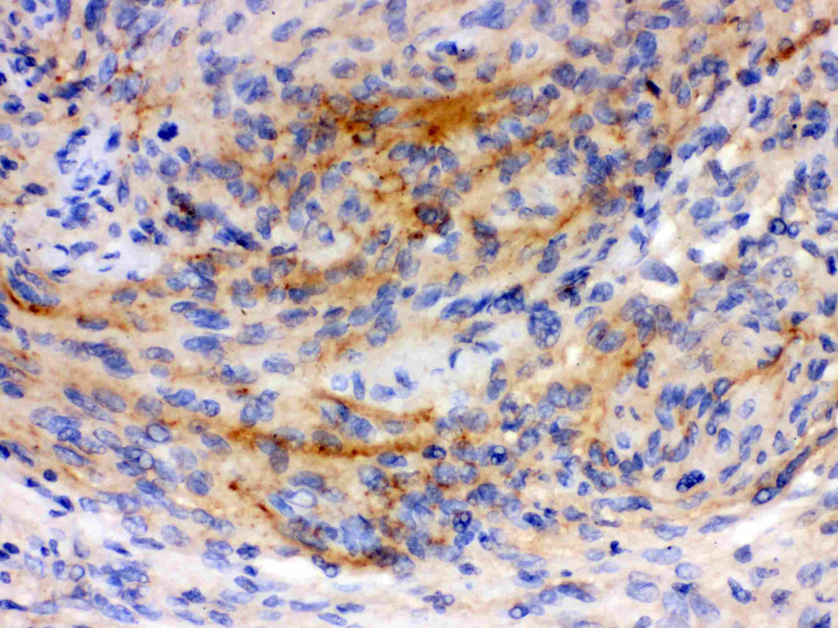
- Immunohistochemistry analysis of GAP43 on paraffin-embedded rat brain tissue. Sample was incubated with GAP43 polyclonal antibody (Product# PA5-79299).
Supportive validation
- Submitted by
- Invitrogen Antibodies (provider)
- Main image
- Experimental details
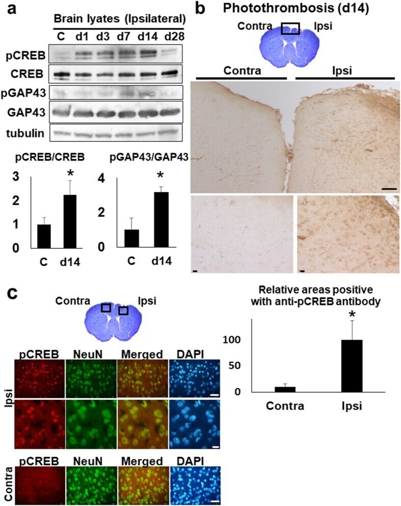
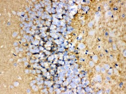
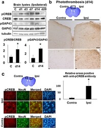
- Fig. 7 The activation of CREB and GAP in the stroke brain. a The western blot analyses with anti-CREB and -GAP43 antibody. The brain lysates (30 mug per each lane) of whole ipsi-lateral cortices at day 1, 3, 7, 14, and 28 were blotted with anti-CREB and anti-GAP43 antibody, respectively. C (control). The lower histogram shows the semi-quantification of band intensities of phosphorylated-CREB and -GAP43 relative to the total CREB and GAP43 protein (C vs. day 14), respectively (* p < 0.05, C vs. day14, Dunnett''s multiple comparison test). The value of control was made 1 for normalization. b The immunohistochemistry with ant-phosphorylated CREB (pCREB) antibody on the brain section, at day 14 post-stroke. The top picture shows the cresyl-violet stained bran section at day 14. Scale bar = 100 mum (upper panels), 10 mum (lower panels). The lower histogram shows the semi-quantification of DAB-stained areas positive with anti- pCREB antibody in ischemic brain sections (Contra- vs. Ipsi-lateral cortex) (* p < 0.05, Contra- vs. Ipsi-lateral, Student''s t-test). The value of ipsilateral brain was made 100 for normalization. Scale bar = 50 mum (upper panels), 10 mum (lower panels). c The immunofluorescence pictures of peri-ischemic regions on the Ipsi- and Contra-lateral cortex at day 14, with anti-phosphorylated CREB (pCREB) and anti-NeuN antibody, respectively. NeuN; Neuron-Specific Nuclear Protein. DAPI; 4'',6-diamidino-2-phenylindole. Scale bar = 50 mum (lower panels)