Explore
 Validate
 Learn
Learn Western blot
 Immunocytochemistry
Antibody data
- Antibody Data
- Antigen structure
- References [4]
- Comments [0]
- Validations
- Western blot [1]
Submit
Validation data
Reference
Comment
Report error
- Product number
- A01868 - Provider product page

- Provider
- Boster Biological Technology
- Product name
- Anti-GAP43 Antibody Picoband™
- Antibody type
- Polyclonal
- Description
- Rabbit IgG polyclonal antibody for GAP43 detection. Tested with WB, IHC-P, IF, FCM in Human;Mouse;Rat.
- Reactivity
- Human, Mouse, Rat
- Host
- Rabbit
- Vial size
- 100μg/vial
- Concentration
- Add 0.2ml of distilled water will yield a concentration of 500ug/ml.
- Storage
- At -20°C for one year. After reconstitution, at 4°C for one month. It can also be aliquoted and stored frozen at -20°C for a longer time. Avoid repeated freezing and thawing.
- Handling
- Add 0.2ml of distilled water will yield a concentration of 500ug/ml.
Submitted references Low-Dose LPS Modulates Microglia/Macrophages Phenotypic Transformation to Amplify Rehabilitation Effects in Chronic Spinal Cord Injured (CSCI) Mice.
Osteopontin enhances the effect of treadmill training and promotes functional recovery after spinal cord injury.
Bone marrow stromal cells improved functional recovery in spinal cord injury rats partly via the Toll-like receptor-4/nuclear factor-κB signaling pathway.
Cograft of neural stem cells and schwann cells overexpressing TrkC and neurotrophin-3 respectively after rat spinal cord transection.
Zhong J, He Y, Zhao Q, Luo H, Zhang Q, Tian Y, Liu Y, Yang C, Yin Y, Yu L, Pan L, Tan B
Molecular neurobiology 2024 Sep;61(9):6484-6500
Molecular neurobiology 2024 Sep;61(9):6484-6500
Osteopontin enhances the effect of treadmill training and promotes functional recovery after spinal cord injury.
Wang Y, Su H, Zhong J, Zhan Z, Zhao Q, Liu Y, Li S, Wang H, Yang C, Yu L, Tan B, Yin Y
Molecular biomedicine 2023 Nov 28;4(1):44
Molecular biomedicine 2023 Nov 28;4(1):44
Bone marrow stromal cells improved functional recovery in spinal cord injury rats partly via the Toll-like receptor-4/nuclear factor-κB signaling pathway.
Bai S, Zhou H, Wu L
Experimental and therapeutic medicine 2019 Jan;17(1):444-448
Experimental and therapeutic medicine 2019 Jan;17(1):444-448
Cograft of neural stem cells and schwann cells overexpressing TrkC and neurotrophin-3 respectively after rat spinal cord transection.
Wang JM, Zeng YS, Wu JL, Li Y, Teng YD
Biomaterials 2011 Oct;32(30):7454-68
Biomaterials 2011 Oct;32(30):7454-68
No comments: Submit comment
Supportive validation
- Submitted by
- Boster Biological Technology (provider)
- Main image

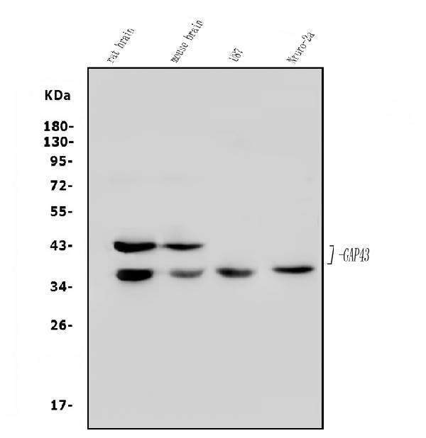
- Experimental details
- Western blot analysis of GAP43 using anti-GAP43 antibody (A01868). Electrophoresis was performed on a 5-20% SDS-PAGE gel at 70V (Stacking gel) / 90V (Resolving gel) for 2-3 hours. The sample well of each lane was loaded with 50ug of sample under reducing conditions. Lane 1: rat brain tissue lysates, Lane 2: mouse brain tissue lysates, Lane 3: human U87 whole cell lysates, Lane 4: mouse Neuro-2a whole cell lysates. After Electrophoresis, proteins were transferred to a Nitrocellulose membrane at 150mA for 50-90 minutes. Blocked the membrane with 5% Non-fat Milk/ TBS for 1.5 hour at RT. The membrane was incubated with rabbit anti-GAP43 antigen affinity purified polyclonal antibody (Catalog # A01868) at 0.5 μg/mL overnight at 4°C, then washed with TBS-0.1%Tween 3 times with 5 minutes each and probed with a goat anti-rabbit IgG-HRP secondary antibody at a dilution of 1:5000 for 1.5 hour at RT. The signal is developed using an Enhanced Chemiluminescent detection (ECL) kit (Catalog # EK1002) with Tanon 5200 system. A specific band was detected for GAP43 at approximately 38-43KD. The expected band size for GAP43 is at 38-43KD.
- Additional image